| Entry | Database: PDB / ID: 4lp9
|
|---|
| Title | Endothiapepsin complexed with Phe-reduced-Tyr peptide. |
|---|
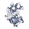
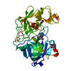
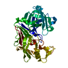
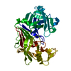
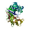
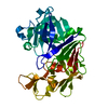
 Components Components | - Endothiapepsin
- Ser-Leu-Phe-His-Phenylalanyl-reduced-peptide-bond-Tyrosyl-Thr-Pro
|
|---|
 Keywords Keywords | hydrolase/hydrolase inhibitor / Aspartic proteinase fold / Proteolysis / HYDROLASE (ACID PROTEINASE) / hydrolase-hydrolase inhibitor complex |
|---|
| Function / homology |  Function and homology information Function and homology information
Aspergillopepsin-like catalytic domain / Eukaryotic aspartyl protease / Aspartic peptidase A1 family / Peptidase family A1 domain / Peptidase family A1 domain profile. / Cathepsin D, subunit A; domain 1 / Acid Proteases / Aspartic peptidase, active site / Eukaryotic and viral aspartyl proteases active site. / Aspartic peptidase domain superfamily ...Aspergillopepsin-like catalytic domain / Eukaryotic aspartyl protease / Aspartic peptidase A1 family / Peptidase family A1 domain / Peptidase family A1 domain profile. / Cathepsin D, subunit A; domain 1 / Acid Proteases / Aspartic peptidase, active site / Eukaryotic and viral aspartyl proteases active site. / Aspartic peptidase domain superfamily / Beta Barrel / Mainly BetaSimilarity search - Domain/homology |
|---|
| Biological species |  Cryphonectria parasitica (chestnut blight fungus) Cryphonectria parasitica (chestnut blight fungus) |
|---|
| Method |  X-RAY DIFFRACTION / SYNCHROTRON / FOURIER SYNTHESIS / Resolution: 1.35 Å X-RAY DIFFRACTION / SYNCHROTRON / FOURIER SYNTHESIS / Resolution: 1.35 Å |
|---|
 Authors Authors | Guo, J. / Cooper, J.B. / Wood, S.P. |
|---|
 Citation Citation | Journal: Acta Crystallogr F Struct Biol Commun / Year: 2014
Title: The structure of endothiapepsin complexed with a Phe-Tyr reduced-bond inhibitor at 1.35 angstrom resolution.
Authors: Guo, J. / Cooper, J.B. / Wood, S.P. |
|---|
| History | | Deposition | Jul 15, 2013 | Deposition site: RCSB / Processing site: RCSB |
|---|
| Revision 1.0 | Jan 15, 2014 | Provider: repository / Type: Initial release |
|---|
| Revision 1.1 | Feb 19, 2014 | Group: Database references |
|---|
| Revision 1.2 | Sep 20, 2023 | Group: Data collection / Database references ...Data collection / Database references / Derived calculations / Refinement description
Category: chem_comp_atom / chem_comp_bond ...chem_comp_atom / chem_comp_bond / database_2 / pdbx_initial_refinement_model / struct_conn / struct_site
Item: _database_2.pdbx_DOI / _database_2.pdbx_database_accession ..._database_2.pdbx_DOI / _database_2.pdbx_database_accession / _struct_conn.pdbx_dist_value / _struct_conn.pdbx_leaving_atom_flag / _struct_conn.ptnr1_auth_comp_id / _struct_conn.ptnr1_auth_seq_id / _struct_conn.ptnr1_label_atom_id / _struct_conn.ptnr1_label_comp_id / _struct_conn.ptnr1_label_seq_id / _struct_conn.ptnr2_auth_comp_id / _struct_conn.ptnr2_auth_seq_id / _struct_conn.ptnr2_label_comp_id / _struct_conn.ptnr2_label_seq_id / _struct_site.pdbx_auth_asym_id / _struct_site.pdbx_auth_comp_id / _struct_site.pdbx_auth_seq_id |
|---|
| Revision 2.0 | Nov 15, 2023 | Group: Atomic model / Data collection / Derived calculations
Category: atom_site / atom_site_anisotrop ...atom_site / atom_site_anisotrop / chem_comp_atom / chem_comp_bond / pdbx_validate_main_chain_plane / pdbx_validate_peptide_omega / pdbx_validate_rmsd_angle / pdbx_validate_torsion / struct_conn
Item: _atom_site.auth_atom_id / _atom_site.label_atom_id ..._atom_site.auth_atom_id / _atom_site.label_atom_id / _atom_site_anisotrop.pdbx_auth_atom_id / _atom_site_anisotrop.pdbx_label_atom_id / _chem_comp_atom.atom_id / _chem_comp_bond.atom_id_1 / _chem_comp_bond.atom_id_2 / _pdbx_validate_peptide_omega.auth_comp_id_1 / _pdbx_validate_peptide_omega.auth_comp_id_2 / _pdbx_validate_peptide_omega.auth_seq_id_1 / _pdbx_validate_peptide_omega.auth_seq_id_2 / _pdbx_validate_peptide_omega.omega / _pdbx_validate_rmsd_angle.angle_deviation / _pdbx_validate_rmsd_angle.angle_standard_deviation / _pdbx_validate_rmsd_angle.angle_target_value / _pdbx_validate_rmsd_angle.angle_value / _pdbx_validate_rmsd_angle.auth_atom_id_1 / _pdbx_validate_rmsd_angle.auth_atom_id_2 / _pdbx_validate_rmsd_angle.auth_atom_id_3 / _pdbx_validate_rmsd_angle.auth_comp_id_1 / _pdbx_validate_rmsd_angle.auth_comp_id_3 / _pdbx_validate_rmsd_angle.auth_seq_id_1 / _pdbx_validate_rmsd_angle.auth_seq_id_3 / _struct_conn.pdbx_leaving_atom_flag / _struct_conn.ptnr1_label_atom_id |
|---|
| Revision 2.1 | Nov 20, 2024 | Group: Structure summary / Category: pdbx_entry_details / pdbx_modification_feature |
|---|
|
|---|
 Movie
Movie Controller


 Open data
 Basic information
 Structure visualization
 Downloads & links
 Other downloads
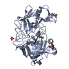


 PDBj
PDBj
 Assembly
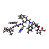
 Type: Peptide-like / Class: Inhibitor / Mass: 998.154 Da / Num. of mol.: 1 / Source method: obtained synthetically
 Mass: 96.063 Da / Num. of mol.: 3 / Source method: obtained synthetically / Formula: SO4
 Mass: 92.094 Da / Num. of mol.: 6 / Source method: obtained synthetically / Formula: C3H8O3
 Mass: 18.015 Da / Num. of mol.: 335 / Source method: isolated from a natural source / Formula: H2O
 Sample preparation
 / Beamline: I02 / Wavelength: 1.0435 Å
 Processing