 Movie
Movie Controller


+ Open data

- Basic information
| Entry | Database: PDB / ID: 4liy | ||||||
|---|---|---|---|---|---|---|---|
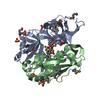
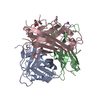
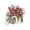
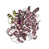
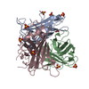
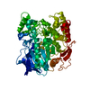
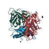
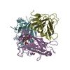
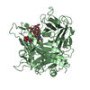
| Title | Structure of the adenovirus 3 knob domain K217E and F224S mutant | ||||||
 Components Components | Fiber protein | ||||||
 Keywords Keywords | VIRAL PROTEIN / adenovirus fibre protein knob domain / viral attachment to host cell / receptor interaction / desmoglein 2 | ||||||
| Function / homology |  Function and homology information Function and homology informationadhesion receptor-mediated virion attachment to host cell / viral capsid / cell adhesion / symbiont entry into host cell / host cell nucleus Similarity search - Function | ||||||
| Biological species |  Human adenovirus 3 Human adenovirus 3 | ||||||
| Method |  X-RAY DIFFRACTION / SYNCHROTRON / MOLECULAR REPLACEMENT / Resolution: 2.1 Å X-RAY DIFFRACTION / SYNCHROTRON / MOLECULAR REPLACEMENT / Resolution: 2.1 Å | ||||||
 Authors Authors | Zubieta, C. / Fender, P. | ||||||
 Citation Citation | Journal: J.Virol. / Year: 2013 Title: Structural and functional studies on the interaction of adenovirus fiber knobs and desmoglein 2 Authors: Wang, H. / Yumul, R. / Cao, H. / Ran, L. / Fan, X. / Richter, M. / Epstein, F. / Gralow, J. / Zubieta, C. / Fender, P. / Lieber, A. | ||||||
| History |
|
- Structure visualization
| Structure viewer | Molecule: MolmilJmol/JSmol |
|---|
- Downloads & links
-Download
| PDBx/mmCIF format | 4liy.cif.gz | 234.3 KB | Display | PDBx/mmCIF format |
|---|---|---|---|---|
| PDB format | pdb4liy.ent.gz | 188.4 KB | Display | PDB format |
| PDBx/mmJSON format | 4liy.json.gz | Tree view | PDBx/mmJSON format | |
| Others |  Other downloads Other downloads |
-Validation report
| Arichive directory | https://data.pdbj.org/pub/pdb/validation_reports/li/4liyftp://data.pdbj.org/pub/pdb/validation_reports/li/4liy | HTTPS FTP |
|---|
-Related structure data
| Related structure data | 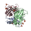 1h7zS S: Starting model for refinement |
|---|---|
| Similar structure data |

-Links
 PDBj
PDBj
- Assembly
| Deposited unit | 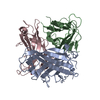
| ||||||||
|---|---|---|---|---|---|---|---|---|---|
| 1 |
| ||||||||
| Unit cell |
|
-Components
| #1: Protein | Mass: 27822.396 Da / Num. of mol.: 3 / Fragment: knob domain, UNP residues 123-319 / Mutation: K217E, F224S Source method: isolated from a genetically manipulated source Source: (gene. exp.) Human adenovirus 3 / Gene: L5 / Plasmid: pQE30 / Production host:  #2: Chemical | ChemComp-SO4 / |   Mass: 96.063 Da / Num. of mol.: 1 / Source method: obtained synthetically / Formula: SO4 Mass: 96.063 Da / Num. of mol.: 1 / Source method: obtained synthetically / Formula: SO4#3: Water | ChemComp-HOH / |  Mass: 18.015 Da / Num. of mol.: 272 / Source method: isolated from a natural source / Formula: H2O Mass: 18.015 Da / Num. of mol.: 272 / Source method: isolated from a natural source / Formula: H2O |
|---|
-Experimental details
-Experiment
| Experiment | Method: X-RAY DIFFRACTION / Number of used crystals: 1 |
|---|
- Sample preparation
| Crystal | Density Matthews: 2.53 Å3/Da / Density % sol: 51.33 % |
|---|---|
| Crystal grow | Temperature: 288 K / Method: vapor diffusion, hanging drop / pH: 9 Details: 1.65M MgSO4, 0.1M TAPS, pH 9, VAPOR DIFFUSION, HANGING DROP, temperature 288K |
-Data collection
| Diffraction | Mean temperature: 100 K |
|---|---|
| Diffraction source | Source: SYNCHROTRON / Site: ESRF  / Beamline: ID14-4 / Wavelength: 0.97239 Å / Beamline: ID14-4 / Wavelength: 0.97239 Å |
| Detector | Type: ADSC QUANTUM 315r / Detector: CCD / Date: May 1, 2013 |
| Radiation | Monochromator: channel cut monochromator / Protocol: SINGLE WAVELENGTH / Monochromatic (M) / Laue (L): M / Scattering type: x-ray |
| Radiation wavelength | Wavelength: 0.97239 Å / Relative weight: 1 |
| Reflection | Resolution: 2.1→48.332 Å / Num. all: 49985 / Num. obs: 49786 / % possible obs: 99.6 % / Observed criterion σ(F): 1.5 / Observed criterion σ(I): 1.5 / Redundancy: 4.4 % / Biso Wilson estimate: 46 Å2 / Rmerge(I) obs: 0.081 / Rsym value: 0.072 / Net I/σ(I): 11.46 |
| Reflection shell | Resolution: 2.1→2.15 Å / Redundancy: 4.3 % / Rmerge(I) obs: 0.632 / Mean I/σ(I) obs: 1.79 / Num. unique all: 3607 / Rsym value: 0.719 / % possible all: 98.6 |
- Processing
| Software |
| |||||||||||||||||||||||||||||||||||||||||||||||||||||||||||||||||||||||||||||||||||||||||||||||||||||||||||||||||||||||||||||||||||||
|---|---|---|---|---|---|---|---|---|---|---|---|---|---|---|---|---|---|---|---|---|---|---|---|---|---|---|---|---|---|---|---|---|---|---|---|---|---|---|---|---|---|---|---|---|---|---|---|---|---|---|---|---|---|---|---|---|---|---|---|---|---|---|---|---|---|---|---|---|---|---|---|---|---|---|---|---|---|---|---|---|---|---|---|---|---|---|---|---|---|---|---|---|---|---|---|---|---|---|---|---|---|---|---|---|---|---|---|---|---|---|---|---|---|---|---|---|---|---|---|---|---|---|---|---|---|---|---|---|---|---|---|---|---|---|
| Refinement | Method to determine structure: MOLECULAR REPLACEMENT Starting model: 1H7Z Resolution: 2.1→48.332 Å / Occupancy max: 1 / Occupancy min: 0.48 / SU ML: 0.22 / Cross valid method: throughput / σ(F): 1.34 / Phase error: 20.22 / Stereochemistry target values: ML
| |||||||||||||||||||||||||||||||||||||||||||||||||||||||||||||||||||||||||||||||||||||||||||||||||||||||||||||||||||||||||||||||||||||
| Solvent computation | Shrinkage radii: 0.9 Å / VDW probe radii: 1.11 Å / Solvent model: FLAT BULK SOLVENT MODEL | |||||||||||||||||||||||||||||||||||||||||||||||||||||||||||||||||||||||||||||||||||||||||||||||||||||||||||||||||||||||||||||||||||||
| Displacement parameters | Biso max: 133.53 Å2 / Biso mean: 49.9725 Å2 / Biso min: 20 Å2 | |||||||||||||||||||||||||||||||||||||||||||||||||||||||||||||||||||||||||||||||||||||||||||||||||||||||||||||||||||||||||||||||||||||
| Refinement step | Cycle: LAST / Resolution: 2.1→48.332 Å
| |||||||||||||||||||||||||||||||||||||||||||||||||||||||||||||||||||||||||||||||||||||||||||||||||||||||||||||||||||||||||||||||||||||
| Refine LS restraints |
| |||||||||||||||||||||||||||||||||||||||||||||||||||||||||||||||||||||||||||||||||||||||||||||||||||||||||||||||||||||||||||||||||||||
| LS refinement shell | Refine-ID: X-RAY DIFFRACTION / Total num. of bins used: 18
| |||||||||||||||||||||||||||||||||||||||||||||||||||||||||||||||||||||||||||||||||||||||||||||||||||||||||||||||||||||||||||||||||||||
| Refinement TLS params. | Method: refined / Origin x: 38.7172 Å / Origin y: -36.5109 Å / Origin z: 12.9515 Å
| |||||||||||||||||||||||||||||||||||||||||||||||||||||||||||||||||||||||||||||||||||||||||||||||||||||||||||||||||||||||||||||||||||||
| Refinement TLS group | Selection details: ALL |
