 Movie
Movie Controller


+ Open data

- Basic information
| Entry | Database: PDB / ID: 4li2 | ||||||
|---|---|---|---|---|---|---|---|
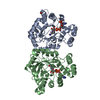
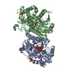
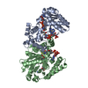
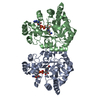
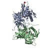
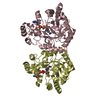
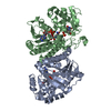
| Title | Crystal Structures of Lgr4 and its complex with R-spondin1 | ||||||
 Components Components |
| ||||||
 Keywords Keywords | Hormone Receptor/Signaling protein / LRR / Hormone Receptor-Signaling protein complex | ||||||
| Function / homology |  Function and homology information Function and homology informationRegulation of FZD by ubiquitination / protein-hormone receptor activity / regulation of receptor internalization / negative regulation of toll-like receptor signaling pathway / bone remodeling / negative regulation of cytokine production / bone mineralization / positive regulation of Wnt signaling pathway / Regulation of FZD by ubiquitination / positive regulation of protein phosphorylation ...Regulation of FZD by ubiquitination / protein-hormone receptor activity / regulation of receptor internalization / negative regulation of toll-like receptor signaling pathway / bone remodeling / negative regulation of cytokine production / bone mineralization / positive regulation of Wnt signaling pathway / Regulation of FZD by ubiquitination / positive regulation of protein phosphorylation / G protein-coupled receptor binding / Wnt signaling pathway / osteoblast differentiation / transmembrane signaling receptor activity / positive regulation of canonical Wnt signaling pathway / rhythmic process / heparin binding / spermatogenesis / G protein-coupled receptor signaling pathway / signaling receptor binding / : / extracellular region / identical protein binding / nucleus / plasma membrane Similarity search - Function | ||||||
| Biological species | Xenopus tropicalis Homo sapiens (human) Homo sapiens (human) | ||||||
| Method |  X-RAY DIFFRACTION / SYNCHROTRON / MOLECULAR REPLACEMENT / Resolution: 3.19 Å X-RAY DIFFRACTION / SYNCHROTRON / MOLECULAR REPLACEMENT / Resolution: 3.19 Å | ||||||
 Authors Authors | Xu, Y. / Rajashankar, K. / Robev, D. | ||||||
 Citation Citation | Journal: Structure / Year: 2013 Title: Crystal structures of lgr4 and its complex with R-spondin1. Authors: Xu, K. / Xu, Y. / Rajashankar, K.R. / Robev, D. / Nikolov, D.B. | ||||||
| History |
|
- Structure visualization
| Structure viewer | Molecule: MolmilJmol/JSmol |
|---|
- Downloads & links
-Download
| PDBx/mmCIF format | 4li2.cif.gz | 114 KB | Display | PDBx/mmCIF format |
|---|---|---|---|---|
| PDB format | pdb4li2.ent.gz | 86.1 KB | Display | PDB format |
| PDBx/mmJSON format | 4li2.json.gz | Tree view | PDBx/mmJSON format | |
| Others |  Other downloads Other downloads |
-Validation report
| Arichive directory | https://data.pdbj.org/pub/pdb/validation_reports/li/4li2ftp://data.pdbj.org/pub/pdb/validation_reports/li/4li2 | HTTPS FTP |
|---|
-Related structure data
| Related structure data | 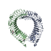 4li1SC S: Starting model for refinement C: citing same article ( |
|---|---|
| Similar structure data |


-Links
 PDBj
PDBj


- Assembly
| Deposited unit | 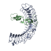
| ||||||||
|---|---|---|---|---|---|---|---|---|---|
| 1 |
| ||||||||
| Unit cell |
| ||||||||
| Details | biological unit is the same as asym. |
-Components
| #1: Protein | Mass: 47542.035 Da / Num. of mol.: 1 / Fragment: EXTRACELLULAR DOMAIN RESIDUES 23-454 / Mutation: C223S Source method: isolated from a genetically manipulated source Source: (gene. exp.) Gene: lgr4 / Plasmid: pCDNA3.1 / Cell line (production host): HEK293 CELLS / Production host: HOMO SAPIENS (human) / References: UniProt: B0BLW3 |
|---|---|
| #2: Protein | Mass: 12153.070 Da / Num. of mol.: 1 / Fragment: FU 1 and FU 2 repeat residues 33-144 Source method: isolated from a genetically manipulated source Source: (gene. exp.) Homo sapiens (human) / Gene: R-spondin1, RSPO1 / Plasmid: pCDNA3.1 / Cell line (production host): HEK293 CELLS / Production host: HOMO SAPIENS (human) / References: UniProt: Q2MKA7 |
| Has protein modification | Y |
-Experimental details
-Experiment
| Experiment | Method: X-RAY DIFFRACTION / Number of used crystals: 1 |
|---|
- Sample preparation
| Crystal | Density Matthews: 2.86 Å3/Da / Density % sol: 57.05 % |
|---|---|
| Crystal grow | Temperature: 298 K / Method: vapor diffusion / pH: 5.7 Details: 22% PEG3350, 200 mM MgCl2 and 100 mM BisTris pH 5.7, vapor diffusion, temperature 298K |
-Data collection
| Diffraction | Mean temperature: 100 K | |||||||||||||||||||||||||||||||||||||||||||||||||||||||||||||||||||||||||||||
|---|---|---|---|---|---|---|---|---|---|---|---|---|---|---|---|---|---|---|---|---|---|---|---|---|---|---|---|---|---|---|---|---|---|---|---|---|---|---|---|---|---|---|---|---|---|---|---|---|---|---|---|---|---|---|---|---|---|---|---|---|---|---|---|---|---|---|---|---|---|---|---|---|---|---|---|---|---|---|
| Diffraction source | Source: SYNCHROTRON / Site: APS  / Beamline: 24-ID-C / Wavelength: 0.9792 Å / Beamline: 24-ID-C / Wavelength: 0.9792 Å | |||||||||||||||||||||||||||||||||||||||||||||||||||||||||||||||||||||||||||||
| Detector | Type: PSI PILATUS 6M / Detector: PIXEL / Date: Feb 8, 2013 | |||||||||||||||||||||||||||||||||||||||||||||||||||||||||||||||||||||||||||||
| Radiation | Protocol: SINGLE WAVELENGTH / Monochromatic (M) / Laue (L): M / Scattering type: x-ray | |||||||||||||||||||||||||||||||||||||||||||||||||||||||||||||||||||||||||||||
| Radiation wavelength | Wavelength: 0.9792 Å / Relative weight: 1 | |||||||||||||||||||||||||||||||||||||||||||||||||||||||||||||||||||||||||||||
| Reflection | Resolution: 3.2→50 Å / Num. obs: 11577 / % possible obs: 98.5 % / Redundancy: 4.4 % / Biso Wilson estimate: 93.49 Å2 / Rmerge(I) obs: 0.087 / Χ2: 0.704 / Net I/σ(I): 5.3 | |||||||||||||||||||||||||||||||||||||||||||||||||||||||||||||||||||||||||||||
| Reflection shell |
|
- Processing
| Software |
| |||||||||||||||||||||||||||||||||||||||||||||||||||||||||||||||
|---|---|---|---|---|---|---|---|---|---|---|---|---|---|---|---|---|---|---|---|---|---|---|---|---|---|---|---|---|---|---|---|---|---|---|---|---|---|---|---|---|---|---|---|---|---|---|---|---|---|---|---|---|---|---|---|---|---|---|---|---|---|---|---|---|
| Refinement | Method to determine structure: MOLECULAR REPLACEMENT Starting model: 4LI1 Resolution: 3.19→47.613 Å / Occupancy max: 1 / Occupancy min: 1 / FOM work R set: 0.6885 / SU ML: 0.46 / σ(F): 1.34 / Phase error: 35.78 / Stereochemistry target values: ML
| |||||||||||||||||||||||||||||||||||||||||||||||||||||||||||||||
| Solvent computation | Shrinkage radii: 0.9 Å / VDW probe radii: 1.11 Å / Solvent model: FLAT BULK SOLVENT MODEL | |||||||||||||||||||||||||||||||||||||||||||||||||||||||||||||||
| Displacement parameters | Biso max: 156.17 Å2 / Biso mean: 71.1696 Å2 / Biso min: 23.53 Å2 | |||||||||||||||||||||||||||||||||||||||||||||||||||||||||||||||
| Refinement step | Cycle: LAST / Resolution: 3.19→47.613 Å
| |||||||||||||||||||||||||||||||||||||||||||||||||||||||||||||||
| Refine LS restraints |
| |||||||||||||||||||||||||||||||||||||||||||||||||||||||||||||||
| LS refinement shell | Refine-ID: X-RAY DIFFRACTION / Total num. of bins used: 8
|
