 Movie
Movie Controller

[English] 日本語
 Yorodumi
- PDB-4ler: Crystal structure of a putative outer membrane protein, probably ... -
+ Open data

- Basic information
| Entry | Database: PDB / ID: 4ler | ||||||
|---|---|---|---|---|---|---|---|
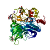
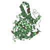
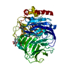
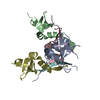
| Title | Crystal structure of a putative outer membrane protein, probably involved in nutrient binding (BVU_1254) from Bacteroides vulgatus ATCC 8482 at 1.42 A resolution | ||||||
 Components Components | Putative outer membrane protein, probably involved in nutrient binding | ||||||
 Keywords Keywords | SUGAR BINDING PROTEIN / SusD homolog / SusD-like_2 family (PF12771) / Structural Genomics / Joint Center for Structural Genomics / JCSG / Protein Structure Initiative / PSI-BIOLOGY | ||||||
| Function / homology |  Function and homology information Function and homology informationSusD-like, tetratrico peptide repeats domain / SusD-like 2 / Starch-binding associating with outer membrane / Serine Threonine Protein Phosphatase 5, Tetratricopeptide repeat - #390 / Four Helix Bundle (Hemerythrin (Met), subunit A) / Serine Threonine Protein Phosphatase 5, Tetratricopeptide repeat / Prokaryotic membrane lipoprotein lipid attachment site profile. / Alpha Horseshoe / Tetratricopeptide-like helical domain superfamily / Up-down Bundle / Mainly Alpha Similarity search - Domain/homology | ||||||
| Biological species |  Bacteroides vulgatus (bacteria) Bacteroides vulgatus (bacteria) | ||||||
| Method |  X-RAY DIFFRACTION / SYNCHROTRON / MAD / Resolution: 1.42 Å X-RAY DIFFRACTION / SYNCHROTRON / MAD / Resolution: 1.42 Å | ||||||
 Authors Authors | Joint Center for Structural Genomics (JCSG) | ||||||
 Citation Citation | Journal: To be published Title: Crystal structure of a putative outer membrane protein, probably involved in nutrient binding (BVU_1254) from Bacteroides vulgatus ATCC 8482 at 1.42 A resolution Authors: Joint Center for Structural Genomics (JCSG) | ||||||
| History |
|
- Structure visualization
| Structure viewer | Molecule: MolmilJmol/JSmol |
|---|
- Downloads & links
-Download
| PDBx/mmCIF format | 4ler.cif.gz | 221.5 KB | Display | PDBx/mmCIF format |
|---|---|---|---|---|
| PDB format | pdb4ler.ent.gz | 175.8 KB | Display | PDB format |
| PDBx/mmJSON format | 4ler.json.gz | Tree view | PDBx/mmJSON format | |
| Others |  Other downloads Other downloads |
-Validation report
| Summary document | 4ler_validation.pdf.gz | 429.9 KB | Display | wwPDB validaton report |
|---|---|---|---|---|
| Full document | 4ler_full_validation.pdf.gz | 431.6 KB | Display | |
| Data in XML | 4ler_validation.xml.gz | 23 KB | Display | |
| Data in CIF | 4ler_validation.cif.gz | 36.5 KB | Display | |
| Arichive directory | https://data.pdbj.org/pub/pdb/validation_reports/le/4lerftp://data.pdbj.org/pub/pdb/validation_reports/le/4ler | HTTPS FTP |
-Related structure data
| Similar structure data | |
|---|---|
| Other databases |

-Links
 PDBj
PDBj


- Assembly
| Deposited unit | 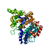
| ||||||||
|---|---|---|---|---|---|---|---|---|---|
| 1 |
| ||||||||
| Unit cell |
| ||||||||
| Details | CRYSTAL PACKING ANALYSIS SUGGESTS THE ASSIGNMENT OF A MONOMER AS THE SIGNIFICANT OLIGOMERIZATION STATE. |
-Components
| #1: Protein | Mass: 53269.320 Da / Num. of mol.: 1 / Fragment: UNP residues 26-493 Source method: isolated from a genetically manipulated source Source: (gene. exp.) Bacteroides vulgatus (bacteria) / Strain: ATCC 8482 / DSM 1447 / NCTC 11154 / Gene: BVU_1254 / Plasmid: SpeedET / Production host: | ||||||
|---|---|---|---|---|---|---|---|
| #2: Chemical |   Mass: 35.453 Da / Num. of mol.: 3 / Source method: obtained synthetically / Formula: Cl Mass: 35.453 Da / Num. of mol.: 3 / Source method: obtained synthetically / Formula: Cl#3: Water | ChemComp-HOH / |  Mass: 18.015 Da / Num. of mol.: 550 / Source method: isolated from a natural source / Formula: H2O Mass: 18.015 Da / Num. of mol.: 550 / Source method: isolated from a natural source / Formula: H2OHas protein modification | Y | Sequence details | THIS CONSTRUCT WAS EXPRESSED WITH A PURIFICATION TAG MGSDKIHHHHHHENLYFQG. THE TAG WAS REMOVED WITH ...THIS CONSTRUCT WAS EXPRESSED WITH A PURIFICATI | |
-Experimental details
-Experiment
| Experiment | Method: X-RAY DIFFRACTION / Number of used crystals: 1 |
|---|
- Sample preparation
| Crystal | Density Matthews: 2.18 Å3/Da / Density % sol: 43.7 % |
|---|---|
| Crystal grow | Temperature: 277 K / Method: vapor diffusion, sitting drop / pH: 6 Details: 1.00M lithium chloride, 20.00% polyethylene glycol 6000, 0.1M MES pH 6.0, NANODROP, VAPOR DIFFUSION, SITTING DROP, temperature 277K |
-Data collection
| Diffraction | Mean temperature: 100 K | |||||||||||||||||||||||||||||||||||||||||||||||||||||||||||||||||||||||||||||
|---|---|---|---|---|---|---|---|---|---|---|---|---|---|---|---|---|---|---|---|---|---|---|---|---|---|---|---|---|---|---|---|---|---|---|---|---|---|---|---|---|---|---|---|---|---|---|---|---|---|---|---|---|---|---|---|---|---|---|---|---|---|---|---|---|---|---|---|---|---|---|---|---|---|---|---|---|---|---|
| Diffraction source | Source: SYNCHROTRON / Site: SSRL  / Beamline: BL11-1 / Wavelength: 0.91837,0.97891,0.97852 / Beamline: BL11-1 / Wavelength: 0.91837,0.97891,0.97852 | |||||||||||||||||||||||||||||||||||||||||||||||||||||||||||||||||||||||||||||
| Detector | Type: DECTRIS PILATUS 6M / Detector: PIXEL / Date: May 23, 2013 Details: Flat mirror (vertical focusing); single crystal Si(111) bent monochromator (horizontal focusing) | |||||||||||||||||||||||||||||||||||||||||||||||||||||||||||||||||||||||||||||
| Radiation | Monochromator: single crystal Si(111) bent / Protocol: MAD / Monochromatic (M) / Laue (L): M / Scattering type: x-ray | |||||||||||||||||||||||||||||||||||||||||||||||||||||||||||||||||||||||||||||
| Radiation wavelength |
| |||||||||||||||||||||||||||||||||||||||||||||||||||||||||||||||||||||||||||||
| Reflection | Resolution: 1.42→29.753 Å / Num. obs: 88866 / % possible obs: 99.3 % / Observed criterion σ(I): -3 / Biso Wilson estimate: 23.315 Å2 / Rmerge(I) obs: 0.064 / Net I/σ(I): 8.43 | |||||||||||||||||||||||||||||||||||||||||||||||||||||||||||||||||||||||||||||
| Reflection shell |
|
-Phasing
| Phasing | Method: MAD |
|---|
- Processing
| Software |
| ||||||||||||||||||||||||||||||||||||||||||||||||||||||||||||||||||||||||||||||||||||||||||||||||||||
|---|---|---|---|---|---|---|---|---|---|---|---|---|---|---|---|---|---|---|---|---|---|---|---|---|---|---|---|---|---|---|---|---|---|---|---|---|---|---|---|---|---|---|---|---|---|---|---|---|---|---|---|---|---|---|---|---|---|---|---|---|---|---|---|---|---|---|---|---|---|---|---|---|---|---|---|---|---|---|---|---|---|---|---|---|---|---|---|---|---|---|---|---|---|---|---|---|---|---|---|---|---|
| Refinement | Method to determine structure: MAD / Resolution: 1.42→29.753 Å / Cor.coef. Fo:Fc: 0.98 / Cor.coef. Fo:Fc free: 0.974 / Occupancy max: 1 / Occupancy min: 0.37 / SU B: 2.189 / SU ML: 0.038 / Cross valid method: THROUGHOUT / σ(F): 0 / ESU R: 0.065 / ESU R Free: 0.056 Stereochemistry target values: MAXIMUM LIKELIHOOD WITH PHASES Details: 1. HYDROGENS HAVE BEEN ADDED IN THE RIDING POSITIONS. 2. A MET-INHIBITION PROTOCOL WAS USED FOR SELENOMETHIONINE INCORPORATION DURING PROTEIN EXPRESSION. THE OCCUPANCY OF THE SE ATOMS IN THE ...Details: 1. HYDROGENS HAVE BEEN ADDED IN THE RIDING POSITIONS. 2. A MET-INHIBITION PROTOCOL WAS USED FOR SELENOMETHIONINE INCORPORATION DURING PROTEIN EXPRESSION. THE OCCUPANCY OF THE SE ATOMS IN THE MSE RESIDUES WAS REDUCED TO 0.75 FOR THE REDUCED SCATTERING POWER DUE TO PARTIAL S-MET INCORPORATION. OTHER REFINEMENT REMARKS: 3. CHLORIDE (CL) IONS FROM PURIFICATION SOLUTIONS HAVE BEEN MODELED. 4. NEGATIVE DIFFERENCE DENSITY ON MANY CARBOXYLATE SIDE CHAINS IS LIKELY THE RESULT OF RADIATION DAMAGE.
| ||||||||||||||||||||||||||||||||||||||||||||||||||||||||||||||||||||||||||||||||||||||||||||||||||||
| Solvent computation | Ion probe radii: 0.8 Å / Shrinkage radii: 0.8 Å / VDW probe radii: 1.4 Å / Solvent model: BABINET MODEL WITH MASK | ||||||||||||||||||||||||||||||||||||||||||||||||||||||||||||||||||||||||||||||||||||||||||||||||||||
| Displacement parameters | Biso max: 59.72 Å2 / Biso mean: 27.0331 Å2 / Biso min: 16.85 Å2
| ||||||||||||||||||||||||||||||||||||||||||||||||||||||||||||||||||||||||||||||||||||||||||||||||||||
| Refinement step | Cycle: LAST / Resolution: 1.42→29.753 Å
| ||||||||||||||||||||||||||||||||||||||||||||||||||||||||||||||||||||||||||||||||||||||||||||||||||||
| Refine LS restraints |
| ||||||||||||||||||||||||||||||||||||||||||||||||||||||||||||||||||||||||||||||||||||||||||||||||||||
| LS refinement shell | Resolution: 1.424→1.46 Å / Total num. of bins used: 20
|
