 Movie
Movie Controller

[English] 日本語
 Yorodumi
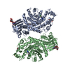
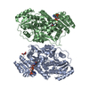
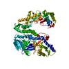
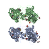
- PDB-4ldy: Crystal structure of the DNA binding domain of the G245A mutant o... -
+ Open data

- Basic information
| Entry | Database: PDB / ID: 4ldy | ||||||
|---|---|---|---|---|---|---|---|
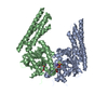
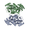
| Title | Crystal structure of the DNA binding domain of the G245A mutant of arabidopsis thaliana auxin reponse factor 1 | ||||||
 Components Components | Auxin response factor 1 | ||||||
 Keywords Keywords | TRANSCRIPTION / Transcription factor / DNA binding / nucleus | ||||||
| Function / homology |  Function and homology information Function and homology informationleaf senescence / cell death / response to auxin / auxin-activated signaling pathway / sequence-specific DNA binding / transcription cis-regulatory region binding / DNA-binding transcription factor activity / negative regulation of DNA-templated transcription / regulation of DNA-templated transcription / DNA binding ...leaf senescence / cell death / response to auxin / auxin-activated signaling pathway / sequence-specific DNA binding / transcription cis-regulatory region binding / DNA-binding transcription factor activity / negative regulation of DNA-templated transcription / regulation of DNA-templated transcription / DNA binding / identical protein binding / nucleus / cytoplasm Similarity search - Function | ||||||
| Biological species |  | ||||||
| Method |  X-RAY DIFFRACTION / SYNCHROTRON / MOLECULAR REPLACEMENT / Resolution: 2.3 Å X-RAY DIFFRACTION / SYNCHROTRON / MOLECULAR REPLACEMENT / Resolution: 2.3 Å | ||||||
 Authors Authors | Boer, D.R. / Freire-Rios, A. / van den Berg, W.M.A. / Weijers, D. / Coll, M. | ||||||
 Citation Citation | Journal: Cell(Cambridge,Mass.) / Year: 2014 Title: Structural Basis for DNA Binding Specificity by the Auxin-Dependent ARF Transcription Factors. Authors: Boer, D.R. / Freire-Rios, A. / van den Berg, W.A. / Saaki, T. / Manfield, I.W. / Kepinski, S. / Lopez-Vidrieo, I. / Franco-Zorrilla, J.M. / de Vries, S.C. / Solano, R. / Weijers, D. / Coll, M. | ||||||
| History |
|
- Structure visualization
| Structure viewer | Molecule: MolmilJmol/JSmol |
|---|
- Downloads & links
-Download
| PDBx/mmCIF format | 4ldy.cif.gz | 276.9 KB | Display | PDBx/mmCIF format |
|---|---|---|---|---|
| PDB format | pdb4ldy.ent.gz | 226.1 KB | Display | PDB format |
| PDBx/mmJSON format | 4ldy.json.gz | Tree view | PDBx/mmJSON format | |
| Others |  Other downloads Other downloads |
-Validation report
| Arichive directory | https://data.pdbj.org/pub/pdb/validation_reports/ld/4ldyftp://data.pdbj.org/pub/pdb/validation_reports/ld/4ldy | HTTPS FTP |
|---|
-Related structure data
| Related structure data |  4lduC 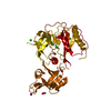 4ldvSC  4ldwC  4ldxC C: citing same article ( S: Starting model for refinement |
|---|---|
| Similar structure data |


-Links
 PDBj
PDBj

- Assembly
| Deposited unit | 
| ||||||||
|---|---|---|---|---|---|---|---|---|---|
| 1 |
| ||||||||
| Unit cell |
|
-Components
| #1: Protein | Mass: 41093.391 Da / Num. of mol.: 2 / Fragment: DNA Binding Domain, UNP residues 1-355 / Mutation: G245A Source method: isolated from a genetically manipulated source Source: (gene. exp.)  #2: Chemical |   Mass: 35.453 Da / Num. of mol.: 2 / Source method: obtained synthetically / Formula: Cl Mass: 35.453 Da / Num. of mol.: 2 / Source method: obtained synthetically / Formula: Cl#3: Water | ChemComp-HOH / |  Mass: 18.015 Da / Num. of mol.: 109 / Source method: isolated from a natural source / Formula: H2O Mass: 18.015 Da / Num. of mol.: 109 / Source method: isolated from a natural source / Formula: H2O |
|---|
-Experimental details
-Experiment
| Experiment | Method: X-RAY DIFFRACTION / Number of used crystals: 1 |
|---|
- Sample preparation
| Crystal | Density Matthews: 2.69 Å3/Da / Density % sol: 54.21 % |
|---|---|
| Crystal grow | Temperature: 293 K / Method: vapor diffusion, sitting drop / pH: 7 Details: 0.75 l of 13 mg/ml Arf1dbd-g245a +0.75 l crystallization buffer (100 mM bis-tris-propane pH 7.0, 0.7 M succinic acid), VAPOR DIFFUSION, SITTING DROP, temperature 293K |
-Data collection
| Diffraction | Mean temperature: 100 K |
|---|---|
| Diffraction source | Source: SYNCHROTRON / Site: ALBA  / Beamline: XALOC / Wavelength: 0.9795 Å / Beamline: XALOC / Wavelength: 0.9795 Å |
| Detector | Type: PSI PILATUS 6M / Detector: PIXEL / Date: Nov 1, 2012 |
| Radiation | Protocol: SINGLE WAVELENGTH / Monochromatic (M) / Laue (L): M / Scattering type: x-ray |
| Radiation wavelength | Wavelength: 0.9795 Å / Relative weight: 1 |
| Reflection | Resolution: 2.3→71.7 Å / Num. obs: 38764 / % possible obs: 99.9 % / Observed criterion σ(I): -3 / Redundancy: 3.1 % / Biso Wilson estimate: 41.1 Å2 / Rmerge(I) obs: 0.102 / Net I/σ(I): 5.9 |
| Reflection shell | Resolution: 2.3→2.42 Å / Redundancy: 3.2 % / Rmerge(I) obs: 0.63 / Mean I/σ(I) obs: 1.8 / Num. unique all: 5641 / % possible all: 100 |
- Processing
| Software |
| ||||||||||||||||||||||||||||||||||||||||||||||||||||||||||||||||||||||||||||||||||||||||||||||||||||||||||||||||||||||||||||||||||||||||||||||||||||||||||||||||||||||||||||||||||||||
|---|---|---|---|---|---|---|---|---|---|---|---|---|---|---|---|---|---|---|---|---|---|---|---|---|---|---|---|---|---|---|---|---|---|---|---|---|---|---|---|---|---|---|---|---|---|---|---|---|---|---|---|---|---|---|---|---|---|---|---|---|---|---|---|---|---|---|---|---|---|---|---|---|---|---|---|---|---|---|---|---|---|---|---|---|---|---|---|---|---|---|---|---|---|---|---|---|---|---|---|---|---|---|---|---|---|---|---|---|---|---|---|---|---|---|---|---|---|---|---|---|---|---|---|---|---|---|---|---|---|---|---|---|---|---|---|---|---|---|---|---|---|---|---|---|---|---|---|---|---|---|---|---|---|---|---|---|---|---|---|---|---|---|---|---|---|---|---|---|---|---|---|---|---|---|---|---|---|---|---|---|---|---|---|
| Refinement | Method to determine structure: MOLECULAR REPLACEMENT Starting model: PDB ENTRY 4LDV Resolution: 2.3→63.91 Å / Cor.coef. Fo:Fc: 0.944 / Cor.coef. Fo:Fc free: 0.917 / SU B: 16.446 / SU ML: 0.194 / Cross valid method: THROUGHOUT / ESU R: 0.29 / ESU R Free: 0.231 / Stereochemistry target values: MAXIMUM LIKELIHOOD / Details: HYDROGENS HAVE BEEN ADDED IN THE RIDING POSITIONS
| ||||||||||||||||||||||||||||||||||||||||||||||||||||||||||||||||||||||||||||||||||||||||||||||||||||||||||||||||||||||||||||||||||||||||||||||||||||||||||||||||||||||||||||||||||||||
| Solvent computation | Ion probe radii: 0.8 Å / Shrinkage radii: 0.8 Å / VDW probe radii: 1.2 Å / Solvent model: MASK | ||||||||||||||||||||||||||||||||||||||||||||||||||||||||||||||||||||||||||||||||||||||||||||||||||||||||||||||||||||||||||||||||||||||||||||||||||||||||||||||||||||||||||||||||||||||
| Displacement parameters | Biso mean: 60.556 Å2
| ||||||||||||||||||||||||||||||||||||||||||||||||||||||||||||||||||||||||||||||||||||||||||||||||||||||||||||||||||||||||||||||||||||||||||||||||||||||||||||||||||||||||||||||||||||||
| Refinement step | Cycle: LAST / Resolution: 2.3→63.91 Å
| ||||||||||||||||||||||||||||||||||||||||||||||||||||||||||||||||||||||||||||||||||||||||||||||||||||||||||||||||||||||||||||||||||||||||||||||||||||||||||||||||||||||||||||||||||||||
| Refine LS restraints |
|