 Movie
Movie Controller

[English] 日本語
 Yorodumi
- PDB-4l9z: Crystal Structure of Rhodobacter sphaeroides malyl-CoA lyase in c... -
+ Open data

- Basic information
| Entry | Database: PDB / ID: 4l9z | ||||||
|---|---|---|---|---|---|---|---|
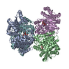
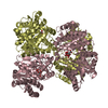
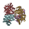
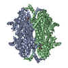
| Title | Crystal Structure of Rhodobacter sphaeroides malyl-CoA lyase in complex with magnesium, oxalate, and CoA | ||||||
 Components Components | Malyl-CoA lyase | ||||||
 Keywords Keywords | LYASE / TIM barrel | ||||||
| Function / homology |  Function and homology information Function and homology informationmalyl-CoA lyase / L-erythro-3-methylmalyl-CoA lyase activity / malyl-CoA lyase activity / (S)-citramalyl-CoA lyase / (S)-citramalyl-CoA lyase activity / oxaloacetate metabolic process / magnesium ion binding / metal ion binding Similarity search - Function | ||||||
| Biological species |  Rhodobacter sphaeroides (bacteria) Rhodobacter sphaeroides (bacteria) | ||||||
| Method |  X-RAY DIFFRACTION / SYNCHROTRON / MOLECULAR REPLACEMENT / Resolution: 2.011 Å X-RAY DIFFRACTION / SYNCHROTRON / MOLECULAR REPLACEMENT / Resolution: 2.011 Å | ||||||
 Authors Authors | Zarzycki, J. / Kerfeld, C.A. | ||||||
 Citation Citation | Journal: Bmc Struct.Biol. / Year: 2013 Title: The crystal structures of the tri-functional Chloroflexus aurantiacus and bi-functional Rhodobacter sphaeroides malyl-CoA lyases and comparison with CitE-like superfamily enzymes and malate synthases. Authors: Zarzycki, J. / Kerfeld, C.A. | ||||||
| History |
|
- Structure visualization
| Structure viewer | Molecule: MolmilJmol/JSmol |
|---|
- Downloads & links
-Download
| PDBx/mmCIF format | 4l9z.cif.gz | 431.5 KB | Display | PDBx/mmCIF format |
|---|---|---|---|---|
| PDB format | pdb4l9z.ent.gz | 349 KB | Display | PDB format |
| PDBx/mmJSON format | 4l9z.json.gz | Tree view | PDBx/mmJSON format | |
| Others |  Other downloads Other downloads |
-Validation report
| Arichive directory | https://data.pdbj.org/pub/pdb/validation_reports/l9/4l9zftp://data.pdbj.org/pub/pdb/validation_reports/l9/4l9z | HTTPS FTP |
|---|
-Related structure data
| Related structure data | 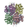 4l7zC 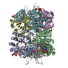 4l80C 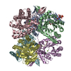 4l9ySC C: citing same article ( S: Starting model for refinement |
|---|---|
| Similar structure data |


-Links
 PDBj
PDBj


- Assembly
| Deposited unit | 
| ||||||||
|---|---|---|---|---|---|---|---|---|---|
| 1 |
| ||||||||
| Unit cell |
|
-Components
| #1: Protein | Mass: 36859.684 Da / Num. of mol.: 6 Source method: isolated from a genetically manipulated source Source: (gene. exp.) Rhodobacter sphaeroides (bacteria) / Strain: ATCC 17023 / 2.4.1 / NCIB 8253 / DSM 158 / Gene: mcl1, RHOS4_03500, RSP_1771 / Plasmid: pET16b / Production host: #2: Chemical | ChemComp-COA /   Mass: 767.534 Da / Num. of mol.: 6 / Source method: obtained synthetically / Formula: C21H36N7O16P3S Mass: 767.534 Da / Num. of mol.: 6 / Source method: obtained synthetically / Formula: C21H36N7O16P3S#3: Chemical | ChemComp-OXL / 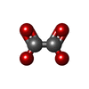  Mass: 88.019 Da / Num. of mol.: 6 / Source method: obtained synthetically / Formula: C2O4 Mass: 88.019 Da / Num. of mol.: 6 / Source method: obtained synthetically / Formula: C2O4#4: Chemical | ChemComp-MG /   Mass: 24.305 Da / Num. of mol.: 6 / Source method: obtained synthetically / Formula: Mg Mass: 24.305 Da / Num. of mol.: 6 / Source method: obtained synthetically / Formula: Mg#5: Water | ChemComp-HOH / |  Mass: 18.015 Da / Num. of mol.: 2526 / Source method: isolated from a natural source / Formula: H2O Mass: 18.015 Da / Num. of mol.: 2526 / Source method: isolated from a natural source / Formula: H2O |
|---|
-Experimental details
-Experiment
| Experiment | Method: X-RAY DIFFRACTION / Number of used crystals: 1 |
|---|
- Sample preparation
| Crystal | Density Matthews: 3.31 Å3/Da / Density % sol: 62.84 % |
|---|---|
| Crystal grow | Temperature: 295 K / Method: vapor diffusion, sitting drop / pH: 7.5 Details: 1 part protein solution (7.5 mg/mL in 20 mM Tris-HCl, pH 7.5, 2 mM magnesium chloride, 100 mM sodium chloride) + 2 parts buffer (0.1 M HEPES/NaOH, pH 7.5, 0.1 M magnesium chloride, 10% w/v ...Details: 1 part protein solution (7.5 mg/mL in 20 mM Tris-HCl, pH 7.5, 2 mM magnesium chloride, 100 mM sodium chloride) + 2 parts buffer (0.1 M HEPES/NaOH, pH 7.5, 0.1 M magnesium chloride, 10% w/v PEG4000) + 1 part substrate solution (20 mM Tris-HCl, pH 7.5, 2 mM magnesium chloride, 100 mM sodium chloride, 20 mM propionyl-CoA, 25 mM sodium oxalate), VAPOR DIFFUSION, SITTING DROP, temperature 295K |
-Data collection
| Diffraction | Mean temperature: 100 K | ||||||||||||||||||||||||||||||||||||||||||||||||||||||||||||||||||||||||||||||||||||||||
|---|---|---|---|---|---|---|---|---|---|---|---|---|---|---|---|---|---|---|---|---|---|---|---|---|---|---|---|---|---|---|---|---|---|---|---|---|---|---|---|---|---|---|---|---|---|---|---|---|---|---|---|---|---|---|---|---|---|---|---|---|---|---|---|---|---|---|---|---|---|---|---|---|---|---|---|---|---|---|---|---|---|---|---|---|---|---|---|---|---|
| Diffraction source | Source: SYNCHROTRON / Site: ALS  / Beamline: 5.0.1 / Wavelength: 0.977408 Å / Beamline: 5.0.1 / Wavelength: 0.977408 Å | ||||||||||||||||||||||||||||||||||||||||||||||||||||||||||||||||||||||||||||||||||||||||
| Detector | Type: ADSC QUANTUM 315r / Detector: CCD / Date: Apr 30, 2012 | ||||||||||||||||||||||||||||||||||||||||||||||||||||||||||||||||||||||||||||||||||||||||
| Radiation | Monochromator: Asymmetric curved crystal Si(220) / Protocol: SINGLE WAVELENGTH / Monochromatic (M) / Laue (L): M / Scattering type: x-ray | ||||||||||||||||||||||||||||||||||||||||||||||||||||||||||||||||||||||||||||||||||||||||
| Radiation wavelength | Wavelength: 0.977408 Å / Relative weight: 1 | ||||||||||||||||||||||||||||||||||||||||||||||||||||||||||||||||||||||||||||||||||||||||
| Reflection | Resolution: 2.011→38.455 Å / Num. all: 175339 / Num. obs: 175339 / % possible obs: 98.3 % / Redundancy: 6.4 % / Rsym value: 0.158 / Net I/σ(I): 11.7 | ||||||||||||||||||||||||||||||||||||||||||||||||||||||||||||||||||||||||||||||||||||||||
| Reflection shell | Diffraction-ID: 1
|
- Processing
| Software |
| |||||||||||||||||||||||||||||||||||||||||||||||||||||||||||||||||||||||||||||||||||||||||||||||||||||||||||||||||||||||||||||||||||||||||||||||||||||||||||||||||||||||||||||||||||||||||||||
|---|---|---|---|---|---|---|---|---|---|---|---|---|---|---|---|---|---|---|---|---|---|---|---|---|---|---|---|---|---|---|---|---|---|---|---|---|---|---|---|---|---|---|---|---|---|---|---|---|---|---|---|---|---|---|---|---|---|---|---|---|---|---|---|---|---|---|---|---|---|---|---|---|---|---|---|---|---|---|---|---|---|---|---|---|---|---|---|---|---|---|---|---|---|---|---|---|---|---|---|---|---|---|---|---|---|---|---|---|---|---|---|---|---|---|---|---|---|---|---|---|---|---|---|---|---|---|---|---|---|---|---|---|---|---|---|---|---|---|---|---|---|---|---|---|---|---|---|---|---|---|---|---|---|---|---|---|---|---|---|---|---|---|---|---|---|---|---|---|---|---|---|---|---|---|---|---|---|---|---|---|---|---|---|---|---|---|---|---|---|---|
| Refinement | Method to determine structure: MOLECULAR REPLACEMENT Starting model: PDB ENTRY 4L9Y Resolution: 2.011→38.455 Å / Occupancy max: 1 / Occupancy min: 0.44 / SU ML: 0.2 / σ(F): 0.86 / Phase error: 19.49 / Stereochemistry target values: ML
| |||||||||||||||||||||||||||||||||||||||||||||||||||||||||||||||||||||||||||||||||||||||||||||||||||||||||||||||||||||||||||||||||||||||||||||||||||||||||||||||||||||||||||||||||||||||||||||
| Solvent computation | Shrinkage radii: 0.9 Å / VDW probe radii: 1.11 Å / Solvent model: FLAT BULK SOLVENT MODEL | |||||||||||||||||||||||||||||||||||||||||||||||||||||||||||||||||||||||||||||||||||||||||||||||||||||||||||||||||||||||||||||||||||||||||||||||||||||||||||||||||||||||||||||||||||||||||||||
| Displacement parameters | Biso max: 80.95 Å2 / Biso mean: 16.0823 Å2 / Biso min: 2.09 Å2 | |||||||||||||||||||||||||||||||||||||||||||||||||||||||||||||||||||||||||||||||||||||||||||||||||||||||||||||||||||||||||||||||||||||||||||||||||||||||||||||||||||||||||||||||||||||||||||||
| Refinement step | Cycle: LAST / Resolution: 2.011→38.455 Å
| |||||||||||||||||||||||||||||||||||||||||||||||||||||||||||||||||||||||||||||||||||||||||||||||||||||||||||||||||||||||||||||||||||||||||||||||||||||||||||||||||||||||||||||||||||||||||||||
| Refine LS restraints |
| |||||||||||||||||||||||||||||||||||||||||||||||||||||||||||||||||||||||||||||||||||||||||||||||||||||||||||||||||||||||||||||||||||||||||||||||||||||||||||||||||||||||||||||||||||||||||||||
| LS refinement shell | Refine-ID: X-RAY DIFFRACTION / Total num. of bins used: 26
|
