 Movie
Movie Controller


+ Open data

- Basic information
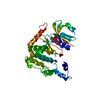
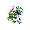
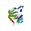
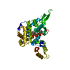
| Entry | Database: PDB / ID: 4l81 | ||||||
|---|---|---|---|---|---|---|---|
| Title | Structure of the SAM-I/IV riboswitch (env87(deltaU92, deltaG93)) | ||||||
 Components Components | SAM-I/IV variant riboswitch aptamer domain | ||||||
 Keywords Keywords | RNA / Riboswitch / Gene regulation / SAM binding | ||||||
| Function / homology | COBALT HEXAMMINE(III) / S-ADENOSYLMETHIONINE / RNA / RNA (> 10) Function and homology information Function and homology information | ||||||
| Method |  X-RAY DIFFRACTION / SYNCHROTRON / MOLECULAR REPLACEMENT / Resolution: 2.95 Å X-RAY DIFFRACTION / SYNCHROTRON / MOLECULAR REPLACEMENT / Resolution: 2.95 Å | ||||||
 Authors Authors | Trausch, J.J. / Reyes, F.E. / Edwards, A.L. / Batey, R.T. | ||||||
 Citation Citation | Journal: Proc.Natl.Acad.Sci.USA / Year: 2014 Title: Structural basis for diversity in the SAM clan of riboswitches. Authors: Trausch, J.J. / Xu, Z. / Edwards, A.L. / Reyes, F.E. / Ross, P.E. / Knight, R. / Batey, R.T. | ||||||
| History |
|
- Structure visualization
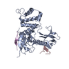
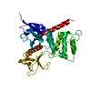
| Structure viewer | Molecule: MolmilJmol/JSmol |
|---|
- Downloads & links
-Download
| PDBx/mmCIF format | 4l81.cif.gz | 65.7 KB | Display | PDBx/mmCIF format |
|---|---|---|---|---|
| PDB format | pdb4l81.ent.gz | 48.6 KB | Display | PDB format |
| PDBx/mmJSON format | 4l81.json.gz | Tree view | PDBx/mmJSON format | |
| Others |  Other downloads Other downloads |
-Validation report
| Arichive directory | https://data.pdbj.org/pub/pdb/validation_reports/l8/4l81ftp://data.pdbj.org/pub/pdb/validation_reports/l8/4l81 | HTTPS FTP |
|---|
-Related structure data
| Related structure data | 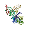 4oquC 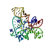 2gisS S: Starting model for refinement C: citing same article ( |
|---|---|
| Similar structure data |


-Links
 PDBj
PDBj


- Assembly
| Deposited unit | 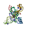
| ||||||||
|---|---|---|---|---|---|---|---|---|---|
| 1 |
| ||||||||
| Unit cell |
|
-Components
| #1: RNA chain | Mass: 31253.779 Da / Num. of mol.: 1 / Fragment: aptamer domain / Source method: obtained synthetically / Details: in vitro T7 RNA polymerase transcript | ||
|---|---|---|---|
| #2: Chemical | ChemComp-SAM /   Mass: 398.437 Da / Num. of mol.: 1 / Source method: obtained synthetically / Formula: C15H22N6O5S Mass: 398.437 Da / Num. of mol.: 1 / Source method: obtained synthetically / Formula: C15H22N6O5S | ||
| #3: Chemical | ChemComp-NCO /   Mass: 161.116 Da / Num. of mol.: 9 / Source method: obtained synthetically / Formula: CoH18N6 Mass: 161.116 Da / Num. of mol.: 9 / Source method: obtained synthetically / Formula: CoH18N6#4: Chemical | ChemComp-MG /   Mass: 24.305 Da / Num. of mol.: 6 / Source method: obtained synthetically / Formula: Mg Mass: 24.305 Da / Num. of mol.: 6 / Source method: obtained synthetically / Formula: Mg |
-Experimental details
-Experiment
| Experiment | Method: X-RAY DIFFRACTION / Number of used crystals: 1 |
|---|
- Sample preparation
| Crystal grow | Temperature: 303 K / Method: vapor diffusion, hanging drop / pH: 8 Details: 50 mM Na-cacodylate, pH 8.0, 40 mM magnesium acetate, 300 mM KCl, 7% 2-methyl-2,4-pentanediol (MPD), 1 mM cobalt hexammine, VAPOR DIFFUSION, HANGING DROP, temperature 303K |
|---|
-Data collection
| Diffraction | Mean temperature: 100 K |
|---|---|
| Diffraction source | Source: SYNCHROTRON / Site: ALS  / Beamline: 8.2.2 / Wavelength: 1 Å / Beamline: 8.2.2 / Wavelength: 1 Å |
| Detector | Type: ADSC QUANTUM 315 / Detector: CCD / Date: Oct 11, 2011 |
| Radiation | Monochromator: Double crystal, Si(111) / Protocol: SINGLE WAVELENGTH / Monochromatic (M) / Laue (L): M / Scattering type: x-ray |
| Radiation wavelength | Wavelength: 1 Å / Relative weight: 1 |
| Reflection | Resolution: 2.95→30 Å / Num. all: 32036 / Num. obs: 31972 / % possible obs: 99.8 % / Observed criterion σ(F): 2 / Observed criterion σ(I): 2 |
| Reflection shell | Resolution: 2.95→3.06 Å / Redundancy: 7.52 % / Rmerge(I) obs: 0.665 / Mean I/σ(I) obs: 2 / % possible all: 99.8 |
- Processing
| Software |
| ||||||||||||||||||||
|---|---|---|---|---|---|---|---|---|---|---|---|---|---|---|---|---|---|---|---|---|---|
| Refinement | Method to determine structure: MOLECULAR REPLACEMENT Starting model: PDB ENTRY 2GIS Resolution: 2.95→30 Å / Cross valid method: THROUGHOUT / σ(F): 2 / Stereochemistry target values: Engh & Huber
| ||||||||||||||||||||
| Refine analyze | Luzzati coordinate error obs: 0.41 Å / Luzzati sigma a obs: 0.63 Å | ||||||||||||||||||||
| Refinement step | Cycle: LAST / Resolution: 2.95→30 Å
| ||||||||||||||||||||
| Refine LS restraints |
|
