 Movie
Movie Controller

[English] 日本語
 Yorodumi
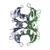
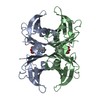
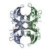
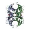
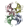
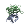
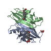
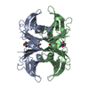
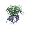
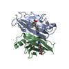
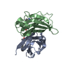
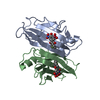
- PDB-4l1t: Transthyretin in complex with (E)-3-(dimethylamino)-5-(4-hydroxy-... -
+ Open data

- Basic information
| Entry | Database: PDB / ID: 4l1t | ||||||
|---|---|---|---|---|---|---|---|
| Title | Transthyretin in complex with (E)-3-(dimethylamino)-5-(4-hydroxy-3,5-dimethylstyryl)benzoic acid | ||||||
 Components Components | Transthyretin | ||||||
 Keywords Keywords | TRANSPORT PROTEIN / Hormone transport protein / Thyroxine / Retinol | ||||||
| Function / homology |  Function and homology information Function and homology informationDefective visual phototransduction due to STRA6 loss of function / negative regulation of glomerular filtration / The canonical retinoid cycle in rods (twilight vision) / purine nucleobase metabolic process / hormone binding / Non-integrin membrane-ECM interactions / phototransduction, visible light / molecular sequestering activity / Retinoid metabolism and transport / retinoid metabolic process ...Defective visual phototransduction due to STRA6 loss of function / negative regulation of glomerular filtration / The canonical retinoid cycle in rods (twilight vision) / purine nucleobase metabolic process / hormone binding / Non-integrin membrane-ECM interactions / phototransduction, visible light / molecular sequestering activity / Retinoid metabolism and transport / retinoid metabolic process / hormone activity / azurophil granule lumen / Amyloid fiber formation / Neutrophil degranulation / protein-containing complex binding / protein-containing complex / : / extracellular exosome / extracellular region / identical protein binding Similarity search - Function | ||||||
| Biological species |  Homo sapiens (human) Homo sapiens (human) | ||||||
| Method |  X-RAY DIFFRACTION / SYNCHROTRON / MOLECULAR REPLACEMENT / Resolution: 1.16 Å X-RAY DIFFRACTION / SYNCHROTRON / MOLECULAR REPLACEMENT / Resolution: 1.16 Å | ||||||
 Authors Authors | Connelly, S. / Wilson, I.A. | ||||||
 Citation Citation | Journal: Biopolymers / Year: 2014 Title: Fluorogenic small molecules requiring reaction with a specific protein to create a fluorescent conjugate for biological imaging-what we know and what we need to learn. Authors: Baranczak, A. / Connelly, S. / Liu, Y. / Choi, S. / Grimster, N.P. / Powers, E.T. / Wilson, I.A. / Kelly, J.W. | ||||||
| History |
|
- Structure visualization
| Structure viewer | Molecule: MolmilJmol/JSmol |
|---|
- Downloads & links
-Download
| PDBx/mmCIF format | 4l1t.cif.gz | 116.5 KB | Display | PDBx/mmCIF format |
|---|---|---|---|---|
| PDB format | pdb4l1t.ent.gz | 90.8 KB | Display | PDB format |
| PDBx/mmJSON format | 4l1t.json.gz | Tree view | PDBx/mmJSON format | |
| Others |  Other downloads Other downloads |
-Validation report
| Arichive directory | https://data.pdbj.org/pub/pdb/validation_reports/l1/4l1tftp://data.pdbj.org/pub/pdb/validation_reports/l1/4l1t | HTTPS FTP |
|---|
-Related structure data
| Related structure data |  4l1sC 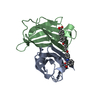 2fbrS S: Starting model for refinement C: citing same article ( |
|---|---|
| Similar structure data |


-Links
 PDBj
PDBj


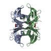
- Assembly
| Deposited unit | 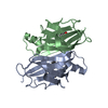
| |||||||||||||||||||||
|---|---|---|---|---|---|---|---|---|---|---|---|---|---|---|---|---|---|---|---|---|---|---|
| 1 | 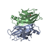
| |||||||||||||||||||||
| Unit cell |
| |||||||||||||||||||||
| Components on special symmetry positions |
|
-Components
| #1: Protein | Mass: 13777.360 Da / Num. of mol.: 2 Source method: isolated from a genetically manipulated source Source: (gene. exp.) Homo sapiens (human) / Gene: TTR, PALB / Plasmid: pmmHA / Production host:  #2: Chemical | 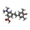  Mass: 311.375 Da / Num. of mol.: 2 / Source method: obtained synthetically / Formula: C19H21NO3 Mass: 311.375 Da / Num. of mol.: 2 / Source method: obtained synthetically / Formula: C19H21NO3#3: Water | ChemComp-HOH / |  Mass: 18.015 Da / Num. of mol.: 179 / Source method: isolated from a natural source / Formula: H2O Mass: 18.015 Da / Num. of mol.: 179 / Source method: isolated from a natural source / Formula: H2O |
|---|
-Experimental details
-Experiment
| Experiment | Method: X-RAY DIFFRACTION / Number of used crystals: 1 |
|---|
- Sample preparation
| Crystal | Density Matthews: 2.07 Å3/Da / Density % sol: 40.55 % |
|---|---|
| Crystal grow | Temperature: 298 K / Method: vapor diffusion, sitting drop / pH: 5.5 Details: The WT-TTR was concentrated to 4 mg/mL in 10 mM NaPi, 100 mM KCl, at pH 7.6 and co-crystallized at room temperature using the vapor diffusion sitting drop method. Crystals were grown from 1. ...Details: The WT-TTR was concentrated to 4 mg/mL in 10 mM NaPi, 100 mM KCl, at pH 7.6 and co-crystallized at room temperature using the vapor diffusion sitting drop method. Crystals were grown from 1.395 M sodium citrate, 3.5% v/v glycerol at pH 5.5. The crystals were frozen using a cryo-protectant solution of 1.395 M sodium citrate, pH 5.5, containing 10% v/v glycerol, VAPOR DIFFUSION, SITTING DROP, temperature 298.0K |
-Data collection
| Diffraction | Mean temperature: 200 K |
|---|---|
| Diffraction source | Source: SYNCHROTRON / Site: SSRL  / Beamline: BL12-2 / Wavelength: 0.9797 Å / Beamline: BL12-2 / Wavelength: 0.9797 Å |
| Detector | Type: PSI PILATUS 6M / Detector: PIXEL / Date: Aug 2, 2012 Details: SINGLE CRYSTAL SI(111) BENT MONOCHROMATOR (HORIZONTAL FOCUSING) |
| Radiation | Monochromator: ASYMMETRIC CUT 4.965 DEGS SIDE SCATTERING BENT CUBE-ROOT I -BEAM SINGLE CRYSTAL Protocol: SINGLE WAVELENGTH / Monochromatic (M) / Laue (L): M / Scattering type: x-ray |
| Radiation wavelength | Wavelength: 0.9797 Å / Relative weight: 1 |
| Reflection | Resolution: 1.16→37.92 Å / Num. obs: 78960 / % possible obs: 98.3 % / Redundancy: 6 % / Biso Wilson estimate: 14 Å2 / Rsym value: 0.034 / Net I/σ(I): 20.3 |
| Reflection shell | Resolution: 1.16→1.22 Å / Redundancy: 5 % / Mean I/σ(I) obs: 2.3 / Num. unique all: 10463 / Rsym value: 0.655 / % possible all: 90.5 |
- Processing
| Software |
| ||||||||||||||||||||||||||||||||||||||||||||||||||||||||||||||||||||||||||||||||||||||||||||||||||||
|---|---|---|---|---|---|---|---|---|---|---|---|---|---|---|---|---|---|---|---|---|---|---|---|---|---|---|---|---|---|---|---|---|---|---|---|---|---|---|---|---|---|---|---|---|---|---|---|---|---|---|---|---|---|---|---|---|---|---|---|---|---|---|---|---|---|---|---|---|---|---|---|---|---|---|---|---|---|---|---|---|---|---|---|---|---|---|---|---|---|---|---|---|---|---|---|---|---|---|---|---|---|
| Refinement | Method to determine structure: MOLECULAR REPLACEMENT Starting model: 2FBR Resolution: 1.16→37.92 Å / Cor.coef. Fo:Fc: 0.973 / Cor.coef. Fo:Fc free: 0.966 / Occupancy max: 1 / Occupancy min: 0.12 / SU B: 1.038 / SU ML: 0.022 / Cross valid method: THROUGHOUT / σ(F): 0 / ESU R: 0.038 / ESU R Free: 0.038 / Stereochemistry target values: MAXIMUM LIKELIHOOD Details: HYDROGENS HAVE BEEN ADDED IN THE RIDING POSITIONS U VALUES : REFINED INDIVIDUALLY
| ||||||||||||||||||||||||||||||||||||||||||||||||||||||||||||||||||||||||||||||||||||||||||||||||||||
| Solvent computation | Ion probe radii: 0.8 Å / Shrinkage radii: 0.8 Å / VDW probe radii: 1.4 Å / Solvent model: MASK | ||||||||||||||||||||||||||||||||||||||||||||||||||||||||||||||||||||||||||||||||||||||||||||||||||||
| Displacement parameters | Biso max: 60.28 Å2 / Biso mean: 18.4022 Å2 / Biso min: 4.46 Å2
| ||||||||||||||||||||||||||||||||||||||||||||||||||||||||||||||||||||||||||||||||||||||||||||||||||||
| Refinement step | Cycle: LAST / Resolution: 1.16→37.92 Å
| ||||||||||||||||||||||||||||||||||||||||||||||||||||||||||||||||||||||||||||||||||||||||||||||||||||
| Refine LS restraints |
| ||||||||||||||||||||||||||||||||||||||||||||||||||||||||||||||||||||||||||||||||||||||||||||||||||||
| LS refinement shell | Resolution: 1.158→1.188 Å / Total num. of bins used: 20
|
