 Movie
Movie Controller

[English] 日本語
 Yorodumi
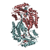
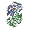
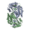
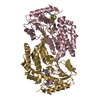
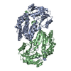
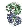
- PDB-4l1o: Crystal structure of human ALDH3A1 with inhibitor 1-{[4-(1,3-benz... -
+ Open data

- Basic information
| Entry | Database: PDB / ID: 4l1o | ||||||
|---|---|---|---|---|---|---|---|
| Title | Crystal structure of human ALDH3A1 with inhibitor 1-{[4-(1,3-benzodioxol-5-ylmethyl)piperazin-1-yl]methyl}-1H-indole-2,3-dione | ||||||
 Components Components | Aldehyde dehydrogenase | ||||||
 Keywords Keywords | OXIDOREDUCTASE/Inhibitor / Catalyzes benzaldehyde / Rossmann fold / Dehydrogenase / NADP+ binding / OXIDOREDUCTASE-Inhibitor complex | ||||||
| Function / homology |  Function and homology information Function and homology informationaldehyde dehydrogenase [NAD(P)+] / 3-chloroallyl aldehyde dehydrogenase activity / benzaldehyde dehydrogenase (NAD+) activity / aldehyde dehydrogenase [NAD(P)+] activity / aldehyde metabolic process / alcohol dehydrogenase (NADP+) activity / aldehyde dehydrogenase (NAD+) activity / Phase I - Functionalization of compounds / xenobiotic metabolic process / lipid metabolic process ...aldehyde dehydrogenase [NAD(P)+] / 3-chloroallyl aldehyde dehydrogenase activity / benzaldehyde dehydrogenase (NAD+) activity / aldehyde dehydrogenase [NAD(P)+] activity / aldehyde metabolic process / alcohol dehydrogenase (NADP+) activity / aldehyde dehydrogenase (NAD+) activity / Phase I - Functionalization of compounds / xenobiotic metabolic process / lipid metabolic process / endoplasmic reticulum / : / plasma membrane / cytoplasm / cytosol Similarity search - Function | ||||||
| Biological species |  Homo sapiens (human) Homo sapiens (human) | ||||||
| Method |  X-RAY DIFFRACTION / MOLECULAR REPLACEMENT / Resolution: 2.3 Å X-RAY DIFFRACTION / MOLECULAR REPLACEMENT / Resolution: 2.3 Å | ||||||
 Authors Authors | Hurley, T.D. / Parajuli, B. | ||||||
 Citation Citation | Journal: J.Med.Chem. / Year: 2014 Title: Development of selective inhibitors for aldehyde dehydrogenases based on substituted indole-2,3-diones. Authors: Kimble-Hill, A.C. / Parajuli, B. / Chen, C.H. / Mochly-Rosen, D. / Hurley, T.D. | ||||||
| History |
|
- Structure visualization
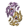
| Structure viewer | Molecule: MolmilJmol/JSmol |
|---|
- Downloads & links
-Download
| PDBx/mmCIF format | 4l1o.cif.gz | 202.1 KB | Display | PDBx/mmCIF format |
|---|---|---|---|---|
| PDB format | pdb4l1o.ent.gz | 158.3 KB | Display | PDB format |
| PDBx/mmJSON format | 4l1o.json.gz | Tree view | PDBx/mmJSON format | |
| Others |  Other downloads Other downloads |
-Validation report
| Arichive directory | https://data.pdbj.org/pub/pdb/validation_reports/l1/4l1oftp://data.pdbj.org/pub/pdb/validation_reports/l1/4l1o | HTTPS FTP |
|---|
-Related structure data
| Related structure data | 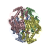 4kwfC 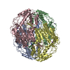 4kwgC 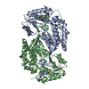 3szaS S: Starting model for refinement C: citing same article ( |
|---|---|
| Similar structure data |


-Links
 PDBj
PDBj

- Assembly
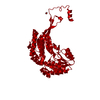
| Deposited unit | 
| ||||||||
|---|---|---|---|---|---|---|---|---|---|
| 1 |
| ||||||||
| Unit cell |
|
-Components
| #1: Protein | Mass: 52245.738 Da / Num. of mol.: 2 Source method: isolated from a genetically manipulated source Source: (gene. exp.) Homo sapiens (human) / Gene: ALDH3, ALDH3A1 / Plasmid: pET28a / Production host:  References: UniProt: P30838, aldehyde dehydrogenase [NAD(P)+] #2: Chemical | 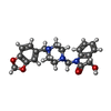  Mass: 381.425 Da / Num. of mol.: 2 / Source method: obtained synthetically / Formula: C21H23N3O4 Mass: 381.425 Da / Num. of mol.: 2 / Source method: obtained synthetically / Formula: C21H23N3O4#3: Chemical | ChemComp-K /   Mass: 39.098 Da / Num. of mol.: 4 / Source method: obtained synthetically / Formula: K Mass: 39.098 Da / Num. of mol.: 4 / Source method: obtained synthetically / Formula: K#4: Chemical |   Mass: 59.044 Da / Num. of mol.: 2 / Source method: obtained synthetically / Formula: C2H3O2 Mass: 59.044 Da / Num. of mol.: 2 / Source method: obtained synthetically / Formula: C2H3O2#5: Water | ChemComp-HOH / |  Mass: 18.015 Da / Num. of mol.: 648 / Source method: isolated from a natural source / Formula: H2O Mass: 18.015 Da / Num. of mol.: 648 / Source method: isolated from a natural source / Formula: H2OHas protein modification | Y | |
|---|
-Experimental details
-Experiment
| Experiment | Method: X-RAY DIFFRACTION / Number of used crystals: 1 |
|---|
- Sample preparation
| Crystal | Density Matthews: 2.16 Å3/Da / Density % sol: 42.93 % |
|---|---|
| Crystal grow | Temperature: 298.15 K / Method: vapor diffusion, sitting drop / pH: 7.5 Details: 0.2 M Potassium Acetate, 20% PEG 3350, (3 microlitres of 3mg/ml of ALDH3A1+ 3 microlitres of mother liquor), pH 7.5, VAPOR DIFFUSION, SITTING DROP, temperature 298.15K |
-Data collection
| Diffraction | Mean temperature: 100 K |
|---|---|
| Diffraction source | Source: ROTATING ANODE / Type: RIGAKU RU200 / Wavelength: 1.5418 Å |
| Detector | Type: RIGAKU RAXIS IV++ / Detector: IMAGE PLATE / Date: May 2, 2011 / Details: OSMIC CONFOCAL BLUE |
| Radiation | Monochromator: OSMIC CONFOCAL BLUE / Protocol: SINGLE WAVELENGTH / Monochromatic (M) / Laue (L): M / Scattering type: x-ray |
| Radiation wavelength | Wavelength: 1.5418 Å / Relative weight: 1 |
| Reflection | Resolution: 2.3→50 Å / Num. obs: 37399 / % possible obs: 90.6 % / Observed criterion σ(F): 0 / Observed criterion σ(I): 0.2 / Redundancy: 3.4 % / Rmerge(I) obs: 0.146 / Rsym value: 0.06 / Net I/σ(I): 16.26 |
| Reflection shell | Resolution: 2.3→2.34 Å / Redundancy: 2.7 % / Rmerge(I) obs: 0.146 / Mean I/σ(I) obs: 6.7 / Num. unique all: 2921 / % possible all: 88.1 |
- Processing
| Software |
| ||||||||||||||||||||||||||||||||||||||||||||||||||||||||||||
|---|---|---|---|---|---|---|---|---|---|---|---|---|---|---|---|---|---|---|---|---|---|---|---|---|---|---|---|---|---|---|---|---|---|---|---|---|---|---|---|---|---|---|---|---|---|---|---|---|---|---|---|---|---|---|---|---|---|---|---|---|---|
| Refinement | Method to determine structure: MOLECULAR REPLACEMENT Starting model: PDB entry 3SZA Resolution: 2.3→47.95 Å / Cor.coef. Fo:Fc: 0.935 / Cor.coef. Fo:Fc free: 0.901 / SU B: 6.435 / SU ML: 0.158 / Cross valid method: THROUGHOUT / ESU R: 0.483 / ESU R Free: 0.245 / Stereochemistry target values: MAXIMUM LIKELIHOOD / Details: HYDROGENS HAVE BEEN ADDED IN THE RIDING POSITIONS
| ||||||||||||||||||||||||||||||||||||||||||||||||||||||||||||
| Solvent computation | Ion probe radii: 0.8 Å / Shrinkage radii: 0.8 Å / VDW probe radii: 1.2 Å / Solvent model: MASK | ||||||||||||||||||||||||||||||||||||||||||||||||||||||||||||
| Displacement parameters | Biso mean: 18.47 Å2
| ||||||||||||||||||||||||||||||||||||||||||||||||||||||||||||
| Refinement step | Cycle: LAST / Resolution: 2.3→47.95 Å
| ||||||||||||||||||||||||||||||||||||||||||||||||||||||||||||
| Refine LS restraints |
| ||||||||||||||||||||||||||||||||||||||||||||||||||||||||||||
| LS refinement shell | Resolution: 2.3→2.359 Å / Total num. of bins used: 20
|
