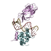
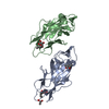
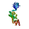
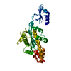
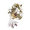
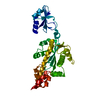
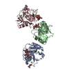
TRANSCRIPTION/DNA / RUNT domain / ETS domain / TRANSCRIPTION-DNA complex
Function / homology
Function and homology information
regulation of hair follicle cell proliferation / SLC-mediated transport of organic cations / positive regulation of progesterone secretion / RUNX1 regulates estrogen receptor mediated transcription / RUNX1 regulates transcription of genes involved in BCR signaling / RUNX1 regulates transcription of genes involved in interleukin signaling / Regulation of RUNX1 Expression and Activity / RUNX1 interacts with co-factors whose precise effect on RUNX1 targets is not known / positive regulation of granulocyte differentiation / RUNX1 and FOXP3 control the development of regulatory T lymphocytes (Tregs) ...regulation of hair follicle cell proliferation / SLC-mediated transport of organic cations / positive regulation of progesterone secretion / RUNX1 regulates estrogen receptor mediated transcription / RUNX1 regulates transcription of genes involved in BCR signaling / RUNX1 regulates transcription of genes involved in interleukin signaling / Regulation of RUNX1 Expression and Activity / RUNX1 interacts with co-factors whose precise effect on RUNX1 targets is not known / positive regulation of granulocyte differentiation / RUNX1 and FOXP3 control the development of regulatory T lymphocytes (Tregs) / RUNX1 regulates transcription of genes involved in differentiation of myeloid cells / core-binding factor complex / positive regulation of cell maturation / positive regulation of CD8-positive, alpha-beta T cell differentiation / myeloid leukocyte differentiation / negative regulation of CD4-positive, alpha-beta T cell differentiation / PML body organization / RUNX1 regulates genes involved in megakaryocyte differentiation and platelet function / negative regulation of granulocyte differentiation / RUNX1 regulates transcription of genes involved in differentiation of HSCs / neuron fate commitment / Estrogen-dependent gene expression / myeloid cell differentiation / myeloid progenitor cell differentiation / definitive hemopoiesis / embryonic hemopoiesis / regulation of T cell anergy / hair follicle morphogenesis / positive regulation of leukocyte adhesion to vascular endothelial cell / negative regulation of cell cycle / regulation of cell differentiation / basement membrane / chondrocyte differentiation / hemopoiesis / behavioral response to pain / regulation of angiogenesis / regulation of signal transduction / positive regulation of blood vessel endothelial cell migration / response to retinoic acid / neuron development / cellular response to transforming growth factor beta stimulus / positive regulation of interleukin-2 production / positive regulation of endothelial cell migration / positive regulation of erythrocyte differentiation / ossification / liver development / transcription corepressor binding / skeletal system development / central nervous system development / cell motility / RNA polymerase II transcription regulatory region sequence-specific DNA binding / promoter-specific chromatin binding / positive regulation of miRNA transcription / Oncogene Induced Senescence / transcription coactivator binding / neuron differentiation / positive regulation of angiogenesis / positive regulation of type II interferon production / sequence-specific double-stranded DNA binding / positive regulation of inflammatory response / in utero embryonic development / transcription by RNA polymerase II / gene expression / DNA-binding transcription activator activity, RNA polymerase II-specific / regulation of apoptotic process / DNA-binding transcription factor binding / nucleic acid binding / RNA polymerase II-specific DNA-binding transcription factor binding / DNA-binding transcription factor activity, RNA polymerase II-specific / cell differentiation / transcription cis-regulatory region binding / immune response / RNA polymerase II cis-regulatory region sequence-specific DNA binding / DNA-binding transcription factor activity / protein heterodimerization activity / negative regulation of cell population proliferation / response to antibiotic / negative regulation of DNA-templated transcription / calcium ion binding / positive regulation of gene expression / regulation of transcription by RNA polymerase II / regulation of DNA-templated transcription / positive regulation of DNA-templated transcription / chromatin / negative regulation of transcription by RNA polymerase II / DNA-templated transcription / protein homodimerization activity / positive regulation of transcription by RNA polymerase II / protein-containing complex / DNA binding / nucleoplasm / ATP binding / identical protein binding / nucleus / cytoplasm Similarity search - Function
Runx, central domain superfamily / Acute myeloid leukemia 1 protein (AML1)/Runt / Runt domain / Runx, C-terminal domain / Runt-related transcription factor RUNX / Runt domain / Runx inhibition domain / Runt domain profile. / Protein C-ets-1, pointed domain / Transforming protein C-ets ...Runx, central domain superfamily / Acute myeloid leukemia 1 protein (AML1)/Runt / Runt domain / Runx, C-terminal domain / Runt-related transcription factor RUNX / Runt domain / Runx inhibition domain / Runt domain profile. / Protein C-ets-1, pointed domain / Transforming protein C-ets / Ets1, N-terminal flanking region of Ets domain / Ets1 N-terminal flanking region of Ets domain / SAM / Pointed domain / Pointed domain / Sterile alpha motif (SAM)/Pointed domain / Pointed (PNT) domain profile. / Ets-domain signature 1. / Immunoglobulin-like - #720 / Ets-domain signature 2. / Ets domain / ETS family / Ets-domain / Ets-domain profile. / erythroblast transformation specific domain / p53/RUNT-type transcription factor, DNA-binding domain superfamily / p53-like transcription factor, DNA-binding / Sterile alpha motif/pointed domain superfamily / Winged helix-like DNA-binding domain superfamily/Winged helix DNA-binding domain / Arc Repressor Mutant, subunit A / Winged helix DNA-binding domain superfamily / Winged helix-like DNA-binding domain superfamily / Immunoglobulin-like / Sandwich / Orthogonal Bundle / Mainly Beta / Mainly Alpha Similarity search - Domain/homology
In the structure databanks used in Yorodumi, some data are registered as the other names, "COVID-19 virus" and "2019-nCoV". Here are the details of the virus and the list of structure data.
Jan 31, 2019. EMDB accession codes are about to change! (news from PDBe EMDB page)
EMDB accession codes are about to change! (news from PDBe EMDB page)
The allocation of 4 digits for EMDB accession codes will soon come to an end. Whilst these codes will remain in use, new EMDB accession codes will include an additional digit and will expand incrementally as the available range of codes is exhausted. The current 4-digit format prefixed with “EMD-” (i.e. EMD-XXXX) will advance to a 5-digit format (i.e. EMD-XXXXX), and so on. It is currently estimated that the 4-digit codes will be depleted around Spring 2019, at which point the 5-digit format will come into force.
The EM Navigator/Yorodumi systems omit the EMD- prefix.
Related info.:Q: What is EMD? / ID/Accession-code notation in Yorodumi/EM Navigator
Yorodumi is a browser for structure data from EMDB, PDB, SASBDB, etc.
This page is also the successor to EM Navigator detail page, and also detail information page/front-end page for Omokage search.
The word "yorodu" (or yorozu) is an old Japanese word meaning "ten thousand". "mi" (miru) is to see.
Related info.:EMDB / PDB / SASBDB / Comparison of 3 databanks / Yorodumi Search / Aug 31, 2016. New EM Navigator & Yorodumi / Yorodumi Papers / Jmol/JSmol / Function and homology information / Changes in new EM Navigator and Yorodumi
 Movie
Movie Controller

 Yorodumi
 Open data
 Basic information
 Components
 Keywords
 Function and homology information
 Homo sapiens (human)
 X-RAY DIFFRACTION /
  Authors
 Citation
 Structure visualization
 Downloads & links
 Other downloads
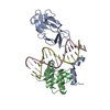


 PDBj
PDBj


 Assembly
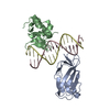

 Mass: 18.015 Da / Num. of mol.: 66 / Source method: isolated from a natural source / Formula: H2O
 Sample preparation
 / Beamline: 24-ID-E / Wavelength: 0.9 Å
 Processing