 Movie
Movie Controller

[English] 日本語
 Yorodumi
- PDB-4l0d: Crystal structure of delta516-525 human cystathionine beta-syntha... -
+ Open data

- Basic information
| Entry | Database: PDB / ID: 4l0d | ||||||
|---|---|---|---|---|---|---|---|
| Title | Crystal structure of delta516-525 human cystathionine beta-synthase containing C-terminal 6xHis-tag | ||||||
 Components Components | Cystathionine beta-synthase | ||||||
 Keywords Keywords | LYASE / CBS domain / homocyteine / cysteine biosynthesis / heme / pyridoxal 5'-phosphate / S-adenosylmethionine / transsulfuration pathway | ||||||
| Function / homology |  Function and homology information Function and homology informationCysteine formation from homocysteine / L-homocysteine catabolic process / modified amino acid binding / cystathionine beta-synthase / cystathionine beta-synthase activity / L-serine catabolic process / : / Metabolism of ingested SeMet, Sec, MeSec into H2Se / : / carbon monoxide binding ...Cysteine formation from homocysteine / L-homocysteine catabolic process / modified amino acid binding / cystathionine beta-synthase / cystathionine beta-synthase activity / L-serine catabolic process / : / Metabolism of ingested SeMet, Sec, MeSec into H2Se / : / carbon monoxide binding / cartilage development involved in endochondral bone morphogenesis / hydrogen sulfide biosynthetic process / homocysteine metabolic process / cerebellum morphogenesis / L-cysteine catabolic process / L-serine metabolic process / L-cysteine biosynthetic process / endochondral ossification / response to folic acid / transsulfuration / nitric oxide binding / : / DNA protection / S-adenosyl-L-methionine binding / nitrite reductase (NO-forming) activity / regulation of JNK cascade / superoxide metabolic process / maternal process involved in female pregnancy / blood vessel remodeling / blood vessel diameter maintenance / oxygen binding / pyridoxal phosphate binding / cellular response to hypoxia / heme binding / ubiquitin protein ligase binding / negative regulation of apoptotic process / enzyme binding / protein homodimerization activity / metal ion binding / identical protein binding / nucleus / cytoplasm / cytosol Similarity search - Function | ||||||
| Biological species |  Homo sapiens (human) Homo sapiens (human) | ||||||
| Method |  X-RAY DIFFRACTION / SYNCHROTRON / MOLECULAR REPLACEMENT / Resolution: 2.97 Å X-RAY DIFFRACTION / SYNCHROTRON / MOLECULAR REPLACEMENT / Resolution: 2.97 Å | ||||||
 Authors Authors | Ereno, J. / Majtan, T. / Oyenarte, I. / Kraus, J.P. / Martinez-Cruz, L.A. | ||||||
 Citation Citation | Journal: Proc.Natl.Acad.Sci.USA / Year: 2013 Title: Structural basis of regulation and oligomerization of human cystathionine beta-synthase, the central enzyme of transsulfuration. Authors: Ereno-Orbea, J. / Majtan, T. / Oyenarte, I. / Kraus, J.P. / Martinez-Cruz, L.A. | ||||||
| History |
|
- Structure visualization
| Structure viewer | Molecule: MolmilJmol/JSmol |
|---|
- Downloads & links
-Download
| PDBx/mmCIF format | 4l0d.cif.gz | 204 KB | Display | PDBx/mmCIF format |
|---|---|---|---|---|
| PDB format | pdb4l0d.ent.gz | 162.2 KB | Display | PDB format |
| PDBx/mmJSON format | 4l0d.json.gz | Tree view | PDBx/mmJSON format | |
| Others |  Other downloads Other downloads |
-Validation report
| Arichive directory | https://data.pdbj.org/pub/pdb/validation_reports/l0/4l0dftp://data.pdbj.org/pub/pdb/validation_reports/l0/4l0d | HTTPS FTP |
|---|
-Related structure data
| Related structure data |  4l27C  4l28C  4l3vC  1jbqS 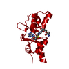 3kpcS S: Starting model for refinement C: citing same article ( |
|---|---|
| Similar structure data |


-Links
 PDBj
PDBj

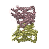
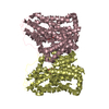
- Assembly
| Deposited unit | 
| ||||||||
|---|---|---|---|---|---|---|---|---|---|
| 1 |
| ||||||||
| Unit cell |
|
-Components
| #1: Protein | Mass: 60605.305 Da / Num. of mol.: 2 / Mutation: P2G Source method: isolated from a genetically manipulated source Source: (gene. exp.) Homo sapiens (human) / Gene: CBS / Production host:  #2: Chemical |   Mass: 247.142 Da / Num. of mol.: 2 / Source method: obtained synthetically / Formula: C8H10NO6P Mass: 247.142 Da / Num. of mol.: 2 / Source method: obtained synthetically / Formula: C8H10NO6P#3: Chemical |   Mass: 616.487 Da / Num. of mol.: 2 / Source method: obtained synthetically / Formula: C34H32FeN4O4 Mass: 616.487 Da / Num. of mol.: 2 / Source method: obtained synthetically / Formula: C34H32FeN4O4 |
|---|
-Experimental details
-Experiment
| Experiment | Method: X-RAY DIFFRACTION / Number of used crystals: 1 |
|---|
- Sample preparation
| Crystal | Density Matthews: 2.96 Å3/Da / Density % sol: 58.41 % |
|---|---|
| Crystal grow | Temperature: 293 K / Method: vapor diffusion, hanging drop / pH: 7.5 Details: 1 M sodium acetate, 0.1 M HEPES, pH 7.5, VAPOR DIFFUSION, HANGING DROP, temperature 293K |
-Data collection
| Diffraction | Mean temperature: 100 K |
|---|---|
| Diffraction source | Source: SYNCHROTRON / Site: ESRF  / Beamline: ID23-2 / Wavelength: 0.9793 Å / Beamline: ID23-2 / Wavelength: 0.9793 Å |
| Detector | Type: MARMOSAIC 225 mm CCD / Detector: CCD / Date: Nov 26, 2011 |
| Radiation | Monochromator: Si(111) / Protocol: SINGLE WAVELENGTH / Monochromatic (M) / Laue (L): M / Scattering type: x-ray |
| Radiation wavelength | Wavelength: 0.9793 Å / Relative weight: 1 |
| Reflection | Resolution: 2.97→53.062 Å / Num. all: 30036 / Num. obs: 29364 / % possible obs: 97.79 % / Observed criterion σ(F): 1 / Observed criterion σ(I): 1 / Redundancy: 3.7 % / Rmerge(I) obs: 0.09 / Net I/σ(I): 9.37 |
| Reflection shell | Resolution: 2.97→3.02 Å / Redundancy: 3.6 % / Rmerge(I) obs: 0.45 / Mean I/σ(I) obs: 2.16 / % possible all: 95.3 |
- Processing
| Software |
| |||||||||||||||||||||||||||||||||||||||||||||||||||||||||||||||||||||||||||||||||||||||||||||||||||||||||||||||||||||||||||||||||||||||||||||||||||
|---|---|---|---|---|---|---|---|---|---|---|---|---|---|---|---|---|---|---|---|---|---|---|---|---|---|---|---|---|---|---|---|---|---|---|---|---|---|---|---|---|---|---|---|---|---|---|---|---|---|---|---|---|---|---|---|---|---|---|---|---|---|---|---|---|---|---|---|---|---|---|---|---|---|---|---|---|---|---|---|---|---|---|---|---|---|---|---|---|---|---|---|---|---|---|---|---|---|---|---|---|---|---|---|---|---|---|---|---|---|---|---|---|---|---|---|---|---|---|---|---|---|---|---|---|---|---|---|---|---|---|---|---|---|---|---|---|---|---|---|---|---|---|---|---|---|---|---|---|
| Refinement | Method to determine structure: MOLECULAR REPLACEMENT Starting model: PDB ENTRIES 1JBQ AND 3KPC Resolution: 2.97→53.062 Å / SU ML: 0.46 / σ(F): 1.09 / Phase error: 33.31 / Stereochemistry target values: ML
| |||||||||||||||||||||||||||||||||||||||||||||||||||||||||||||||||||||||||||||||||||||||||||||||||||||||||||||||||||||||||||||||||||||||||||||||||||
| Solvent computation | Shrinkage radii: 0.9 Å / VDW probe radii: 1.11 Å / Solvent model: FLAT BULK SOLVENT MODEL | |||||||||||||||||||||||||||||||||||||||||||||||||||||||||||||||||||||||||||||||||||||||||||||||||||||||||||||||||||||||||||||||||||||||||||||||||||
| Refinement step | Cycle: LAST / Resolution: 2.97→53.062 Å
| |||||||||||||||||||||||||||||||||||||||||||||||||||||||||||||||||||||||||||||||||||||||||||||||||||||||||||||||||||||||||||||||||||||||||||||||||||
| Refine LS restraints |
| |||||||||||||||||||||||||||||||||||||||||||||||||||||||||||||||||||||||||||||||||||||||||||||||||||||||||||||||||||||||||||||||||||||||||||||||||||
| LS refinement shell |
|
