 Movie
Movie Controller

[English] 日本語
 Yorodumi
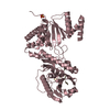
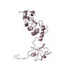
- PDB-4kzs: Crystal structure of the secreted protein HP1454 from the human p... -
+ Open data

- Basic information
| Entry | Database: PDB / ID: 4kzs | ||||||
|---|---|---|---|---|---|---|---|
| Title | Crystal structure of the secreted protein HP1454 from the human pathogen Helicobacter pylori | ||||||
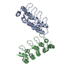
 Components Components | LPP20 lipofamily protein | ||||||
 Keywords Keywords | UNKNOWN FUNCTION / Helicobacter pylori / secreted proteins / outer membrane / Tol-Pal system / Three-helix bundle | ||||||
| Function / homology |  Function and homology information Function and homology informationDouble Stranded RNA Binding Domain - #710 / Helix Hairpins - #1870 / : / : / LPP20 lipofamily protein, C-terminal domain / LPP20 lipofamily protein, middle domain / Acetamidase/Formamidase-like domains / Lipoprotein LPP20-like / LPP20 lipoprotein / Endonuclease I-creI ...Double Stranded RNA Binding Domain - #710 / Helix Hairpins - #1870 / : / : / LPP20 lipofamily protein, C-terminal domain / LPP20 lipofamily protein, middle domain / Acetamidase/Formamidase-like domains / Lipoprotein LPP20-like / LPP20 lipoprotein / Endonuclease I-creI / Helix Hairpins / Double Stranded RNA Binding Domain / Helix non-globular / Special / Roll / 2-Layer Sandwich / Alpha Beta Similarity search - Domain/homology | ||||||
| Biological species |  Helicobacter pylori R046Wa (bacteria) Helicobacter pylori R046Wa (bacteria) | ||||||
| Method |  X-RAY DIFFRACTION / SYNCHROTRON / MAD / Resolution: 2.7 Å X-RAY DIFFRACTION / SYNCHROTRON / MAD / Resolution: 2.7 Å | ||||||
 Authors Authors | Quarantini, S. / Cendron, L. / Fischer, W. / Zanotti, G. | ||||||
 Citation Citation | Journal: Proteins / Year: 2014 Title: Crystal structure of the secreted protein HP1454 from the human pathogen Helicobacter pylori. Authors: Quarantini, S. / Cendron, L. / Zanotti, G. | ||||||
| History |
|
- Structure visualization
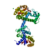
| Structure viewer | Molecule: MolmilJmol/JSmol |
|---|
- Downloads & links
-Download
| PDBx/mmCIF format | 4kzs.cif.gz | 63.8 KB | Display | PDBx/mmCIF format |
|---|---|---|---|---|
| PDB format | pdb4kzs.ent.gz | 46.7 KB | Display | PDB format |
| PDBx/mmJSON format | 4kzs.json.gz | Tree view | PDBx/mmJSON format | |
| Others |  Other downloads Other downloads |
-Validation report
| Arichive directory | https://data.pdbj.org/pub/pdb/validation_reports/kz/4kzsftp://data.pdbj.org/pub/pdb/validation_reports/kz/4kzs | HTTPS FTP |
|---|
-Related structure data
| Similar structure data |
|---|
-Links
 PDBj
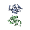
PDBj- Assembly
| Deposited unit | 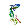
| |||||||||
|---|---|---|---|---|---|---|---|---|---|---|
| 1 |
| |||||||||
| Unit cell |
| |||||||||
| Components on special symmetry positions |
|
-Components
| #1: Protein | Mass: 32019.309 Da / Num. of mol.: 1 / Mutation: V10M Source method: isolated from a genetically manipulated source Source: (gene. exp.) Helicobacter pylori R046Wa (bacteria) / Strain: CCUG 17874 / Gene: hp1454, OUO_1490 / Plasmid: HP1454-pET151 / Production host: |
|---|---|
| #2: Chemical | ChemComp-1PE /   Mass: 238.278 Da / Num. of mol.: 1 / Source method: obtained synthetically / Formula: C10H22O6 / Comment: precipitant*YM Mass: 238.278 Da / Num. of mol.: 1 / Source method: obtained synthetically / Formula: C10H22O6 / Comment: precipitant*YM |
| #3: Water | ChemComp-HOH /  Mass: 18.015 Da / Num. of mol.: 35 / Source method: isolated from a natural source / Formula: H2O Mass: 18.015 Da / Num. of mol.: 35 / Source method: isolated from a natural source / Formula: H2O |
-Experimental details
-Experiment
| Experiment | Method: X-RAY DIFFRACTION / Number of used crystals: 1 |
|---|
- Sample preparation
| Crystal | Density Matthews: 3.33 Å3/Da / Density % sol: 63.06 % |
|---|---|
| Crystal grow | Temperature: 298 K / Method: vapor diffusion / pH: 4.6 Details: 0.1 M Sodium Acetate, and 2.0 M Ammonium Sulfate, pH 4.6, VAPOR DIFFUSION, temperature 298K |
-Data collection
| Diffraction | Mean temperature: 100 K | |||||||||
|---|---|---|---|---|---|---|---|---|---|---|
| Diffraction source | Source: SYNCHROTRON / Site: ESRF  / Beamline: BM14 / Wavelength: 0.97887, 0.97826 / Beamline: BM14 / Wavelength: 0.97887, 0.97826 | |||||||||
| Detector | Type: MARMOSAIC 225 mm CCD / Detector: CCD / Date: May 5, 2011 | |||||||||
| Radiation | Protocol: MAD / Monochromatic (M) / Laue (L): M / Scattering type: x-ray | |||||||||
| Radiation wavelength |
| |||||||||
| Reflection | Resolution: 2.7→60.54 Å / Num. all: 12784 / Num. obs: 12784 / % possible obs: 99.4 % / Observed criterion σ(F): 0 / Observed criterion σ(I): 0 / Redundancy: 9.1 % / Rmerge(I) obs: 0.092 / Net I/σ(I): 14.3 | |||||||||
| Reflection shell | Resolution: 2.7→2.85 Å / Redundancy: 9.5 % / Rmerge(I) obs: 0.609 / Mean I/σ(I) obs: 3.3 / Num. unique all: 1854 / % possible all: 100 |
- Processing
| Software |
| ||||||||||||||||||||||||||||||||||||||||||||||||||||||||||||||||||||||||||||||||||||||||||||||||||||||||||||||||||||||||||||||||||||||||||||||||||||||||||||||||||||||||||
|---|---|---|---|---|---|---|---|---|---|---|---|---|---|---|---|---|---|---|---|---|---|---|---|---|---|---|---|---|---|---|---|---|---|---|---|---|---|---|---|---|---|---|---|---|---|---|---|---|---|---|---|---|---|---|---|---|---|---|---|---|---|---|---|---|---|---|---|---|---|---|---|---|---|---|---|---|---|---|---|---|---|---|---|---|---|---|---|---|---|---|---|---|---|---|---|---|---|---|---|---|---|---|---|---|---|---|---|---|---|---|---|---|---|---|---|---|---|---|---|---|---|---|---|---|---|---|---|---|---|---|---|---|---|---|---|---|---|---|---|---|---|---|---|---|---|---|---|---|---|---|---|---|---|---|---|---|---|---|---|---|---|---|---|---|---|---|---|---|---|---|---|
| Refinement | Method to determine structure: MAD / Resolution: 2.7→33.44 Å / Cor.coef. Fo:Fc: 0.925 / Cor.coef. Fo:Fc free: 0.859 / SU B: 13.2 / SU ML: 0.272 / Cross valid method: THROUGHOUT / ESU R: 0.464 / ESU R Free: 0.338 / Stereochemistry target values: MAXIMUM LIKELIHOOD / Details: HYDROGENS HAVE BEEN USED IF PRESENT IN THE INPUT
| ||||||||||||||||||||||||||||||||||||||||||||||||||||||||||||||||||||||||||||||||||||||||||||||||||||||||||||||||||||||||||||||||||||||||||||||||||||||||||||||||||||||||||
| Solvent computation | Ion probe radii: 0.8 Å / Shrinkage radii: 0.8 Å / VDW probe radii: 1.2 Å / Solvent model: BABINET MODEL WITH MASK | ||||||||||||||||||||||||||||||||||||||||||||||||||||||||||||||||||||||||||||||||||||||||||||||||||||||||||||||||||||||||||||||||||||||||||||||||||||||||||||||||||||||||||
| Displacement parameters | Biso mean: 85.21 Å2
| ||||||||||||||||||||||||||||||||||||||||||||||||||||||||||||||||||||||||||||||||||||||||||||||||||||||||||||||||||||||||||||||||||||||||||||||||||||||||||||||||||||||||||
| Refinement step | Cycle: LAST / Resolution: 2.7→33.44 Å
| ||||||||||||||||||||||||||||||||||||||||||||||||||||||||||||||||||||||||||||||||||||||||||||||||||||||||||||||||||||||||||||||||||||||||||||||||||||||||||||||||||||||||||
| Refine LS restraints |
| ||||||||||||||||||||||||||||||||||||||||||||||||||||||||||||||||||||||||||||||||||||||||||||||||||||||||||||||||||||||||||||||||||||||||||||||||||||||||||||||||||||||||||
| LS refinement shell | Resolution: 2.7→2.77 Å / Total num. of bins used: 20
|
