 Movie
Movie Controller

[English] 日本語
 Yorodumi
- PDB-4kyn: Crystal structure of odorant binding protein 48 from Anopheles ga... -
+ Open data

- Basic information
| Entry | Database: PDB / ID: 4kyn | ||||||
|---|---|---|---|---|---|---|---|
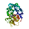
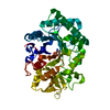
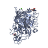
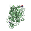
| Title | Crystal structure of odorant binding protein 48 from Anopheles gambiae at 3.3 Angstrom resolution | ||||||
 Components Components | Odorant binding protein-8 | ||||||
 Keywords Keywords | TRANSPORT PROTEIN / insect odorant binding protein / OBP48 / olfaction / Anopheles gambiae | ||||||
| Function / homology | Recoverin; domain 1 - #270 / : / : / AgamOBP47-like / Recoverin; domain 1 / Orthogonal Bundle / extracellular region / Mainly Alpha / Odorant binding protein-8 Function and homology information Function and homology information | ||||||
| Biological species |  | ||||||
| Method |  X-RAY DIFFRACTION / SYNCHROTRON / MOLECULAR REPLACEMENT / molecular replacement / Resolution: 3.3 Å X-RAY DIFFRACTION / SYNCHROTRON / MOLECULAR REPLACEMENT / molecular replacement / Resolution: 3.3 Å | ||||||
 Authors Authors | Tsitsanou, K.E. / Drakou, C.E. / Zographos, S.E. | ||||||
 Citation Citation | Journal: J.Biol.Chem. / Year: 2013 Title: Crystal and Solution Studies of the "Plus-C" Odorant-binding Protein 48 from Anopheles gambiae: CONTROL OF BINDING SPECIFICITY THROUGH THREE-DIMENSIONAL DOMAIN SWAPPING. Authors: Tsitsanou, K.E. / Drakou, C.E. / Thireou, T. / Vitlin Gruber, A. / Kythreoti, G. / Azem, A. / Fessas, D. / Eliopoulos, E. / Iatrou, K. / Zographos, S.E. | ||||||
| History |
|
- Structure visualization
| Structure viewer | Molecule: MolmilJmol/JSmol |
|---|
- Downloads & links
-Download
| PDBx/mmCIF format | 4kyn.cif.gz | 135.8 KB | Display | PDBx/mmCIF format |
|---|---|---|---|---|
| PDB format | pdb4kyn.ent.gz | 109 KB | Display | PDB format |
| PDBx/mmJSON format | 4kyn.json.gz | Tree view | PDBx/mmJSON format | |
| Others |  Other downloads Other downloads |
-Validation report
| Arichive directory | https://data.pdbj.org/pub/pdb/validation_reports/ky/4kynftp://data.pdbj.org/pub/pdb/validation_reports/ky/4kyn | HTTPS FTP |
|---|
-Related structure data
| Related structure data |  4ij7SC S: Starting model for refinement C: citing same article ( |
|---|---|
| Similar structure data |


-Links
 PDBj
PDBj- Assembly
| Deposited unit | 
| ||||||||
|---|---|---|---|---|---|---|---|---|---|
| 1 | 
| ||||||||
| 2 | 
| ||||||||
| Unit cell |
|
-Components
| #1: Protein | Mass: 19041.273 Da / Num. of mol.: 4 / Fragment: UNP residues 29-200 Source method: isolated from a genetically manipulated source Source: (gene. exp.) Gene: agamobp48, OBP-8 / Plasmid: pET-22b(+) / Production host:  Has protein modification | Y | |
|---|
-Experimental details
-Experiment
| Experiment | Method: X-RAY DIFFRACTION / Number of used crystals: 1 |
|---|
- Sample preparation
| Crystal | Density Matthews: 2.62 Å3/Da / Density % sol: 53.03 % |
|---|---|
| Crystal grow | Temperature: 293 K / Method: vapor diffusion, sitting drop / pH: 7 Details: 4.0 M sodium formate, pH 7, VAPOR DIFFUSION, SITTING DROP, temperature 293K |
-Data collection
| Diffraction | Mean temperature: 100 K |
|---|---|
| Diffraction source | Source: SYNCHROTRON / Site: MAX II  / Beamline: I911-3 / Wavelength: 1 Å / Beamline: I911-3 / Wavelength: 1 Å |
| Detector | Type: MARMOSAIC 225 mm CCD / Detector: CCD / Date: Feb 23, 2013 Details: 1st mirror: Rh-coated Si mirror, bent for vertical collimation; 2nd mirror: Rh-coated toroidal Si mirror |
| Radiation | Monochromator: double crystal Si(111), first crystal indirectly water-cooled Protocol: SINGLE WAVELENGTH / Monochromatic (M) / Laue (L): M / Scattering type: x-ray |
| Radiation wavelength | Wavelength: 1 Å / Relative weight: 1 |
| Reflection twin | Operator: h,-h-k,-l / Fraction: 0.5 |
| Reflection | Resolution: 3.3→45.73 Å / Num. all: 11617 / Num. obs: 11606 / % possible obs: 99.9 % / Observed criterion σ(F): 0 / Observed criterion σ(I): 0 / Redundancy: 4 % / Biso Wilson estimate: 52.01 Å2 / Rmerge(I) obs: 0.112 / Rsym value: 0.112 / Net I/σ(I): 6.7 |
| Reflection shell | Resolution: 3.3→3.48 Å / Redundancy: 4 % / Rmerge(I) obs: 0.477 / Mean I/σ(I) obs: 2.3 / Num. unique all: 1661 / Rsym value: 0.477 / % possible all: 100 |
-Phasing
| Phasing | Method: molecular replacement |
|---|
- Processing
| Software |
| |||||||||||||||||||||||||||||||||||
|---|---|---|---|---|---|---|---|---|---|---|---|---|---|---|---|---|---|---|---|---|---|---|---|---|---|---|---|---|---|---|---|---|---|---|---|---|
| Refinement | Method to determine structure: MOLECULAR REPLACEMENT Starting model: PDB ENTRY 4IJ7 Resolution: 3.3→44.967 Å / Occupancy max: 1 / Occupancy min: 1 / σ(F): 1.98 / Phase error: 23.9 / Stereochemistry target values: TWIN_LSQ_F
| |||||||||||||||||||||||||||||||||||
| Solvent computation | Shrinkage radii: 0.9 Å / VDW probe radii: 1.11 Å / Solvent model: FLAT BULK SOLVENT MODEL | |||||||||||||||||||||||||||||||||||
| Displacement parameters | Biso max: 119.83 Å2 / Biso mean: 53.7713 Å2 / Biso min: 19.17 Å2 | |||||||||||||||||||||||||||||||||||
| Refine analyze | Luzzati coordinate error obs: 0.718 Å | |||||||||||||||||||||||||||||||||||
| Refinement step | Cycle: LAST / Resolution: 3.3→44.967 Å
| |||||||||||||||||||||||||||||||||||
| Refine LS restraints |
| |||||||||||||||||||||||||||||||||||
| LS refinement shell | Refine-ID: X-RAY DIFFRACTION / Total num. of bins used: 4
|
