 Movie
Movie Controller

[English] 日本語
 Yorodumi
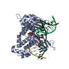
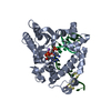
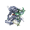
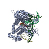
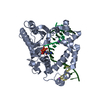
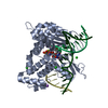
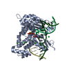
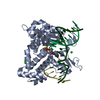
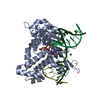
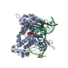
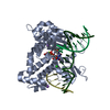
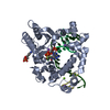
- PDB-4kls: DNA polymerase beta mismatched reactant complex with Mn2+, 10 min -
+ Open data

- Basic information
| Entry | Database: PDB / ID: 4kls | ||||||
|---|---|---|---|---|---|---|---|
| Title | DNA polymerase beta mismatched reactant complex with Mn2+, 10 min | ||||||
 Components Components |
| ||||||
 Keywords Keywords | TRANSFERASE / LYASE/DNA / DNA polymerase / LYASE-DNA complex | ||||||
| Function / homology |  Function and homology information Function and homology informationResolution of AP sites via the single-nucleotide replacement pathway / immunoglobulin heavy chain V-D-J recombination / Resolution of AP sites via the multiple-nucleotide patch replacement pathway / Abasic sugar-phosphate removal via the single-nucleotide replacement pathway / APEX1-Independent Resolution of AP Sites via the Single Nucleotide Replacement Pathway / Lyases; Carbon-oxygen lyases; Other carbon-oxygen lyases / homeostasis of number of cells / pyrimidine dimer repair / POLB-Dependent Long Patch Base Excision Repair / PCNA-Dependent Long Patch Base Excision Repair ...Resolution of AP sites via the single-nucleotide replacement pathway / immunoglobulin heavy chain V-D-J recombination / Resolution of AP sites via the multiple-nucleotide patch replacement pathway / Abasic sugar-phosphate removal via the single-nucleotide replacement pathway / APEX1-Independent Resolution of AP Sites via the Single Nucleotide Replacement Pathway / Lyases; Carbon-oxygen lyases; Other carbon-oxygen lyases / homeostasis of number of cells / pyrimidine dimer repair / POLB-Dependent Long Patch Base Excision Repair / PCNA-Dependent Long Patch Base Excision Repair / 5'-deoxyribose-5-phosphate lyase activity / response to hyperoxia / lymph node development / spleen development / salivary gland morphogenesis / somatic hypermutation of immunoglobulin genes / base-excision repair, gap-filling / DNA-(apurinic or apyrimidinic site) endonuclease activity / class I DNA-(apurinic or apyrimidinic site) endonuclease activity / DNA-(apurinic or apyrimidinic site) lyase / response to gamma radiation / spindle microtubule / base-excision repair / DNA-templated DNA replication / double-strand break repair via nonhomologous end joining / intrinsic apoptotic signaling pathway in response to DNA damage / neuron apoptotic process / in utero embryonic development / DNA-directed DNA polymerase / microtubule binding / damaged DNA binding / microtubule / response to ethanol / DNA-directed DNA polymerase activity / lyase activity / Ub-specific processing proteases / inflammatory response / DNA repair / DNA damage response / enzyme binding / protein-containing complex / nucleoplasm / metal ion binding / nucleus / cytoplasm Similarity search - Function | ||||||
| Biological species |  Homo sapiens (human) Homo sapiens (human) | ||||||
| Method |  X-RAY DIFFRACTION / MOLECULAR REPLACEMENT / Resolution: 1.978 Å X-RAY DIFFRACTION / MOLECULAR REPLACEMENT / Resolution: 1.978 Å | ||||||
 Authors Authors | Freudenthal, B.D. / Beard, W.A. / Shock, D.D. / Wilson, S.H. | ||||||
 Citation Citation | Journal: Cell(Cambridge,Mass.) / Year: 2013 Title: Observing a DNA polymerase choose right from wrong. Authors: Freudenthal, B.D. / Beard, W.A. / Shock, D.D. / Wilson, S.H. | ||||||
| History |
|
- Structure visualization
| Structure viewer | Molecule: MolmilJmol/JSmol |
|---|
- Downloads & links
-Download
| PDBx/mmCIF format | 4kls.cif.gz | 109.9 KB | Display | PDBx/mmCIF format |
|---|---|---|---|---|
| PDB format | pdb4kls.ent.gz | 78.2 KB | Display | PDB format |
| PDBx/mmJSON format | 4kls.json.gz | Tree view | PDBx/mmJSON format | |
| Others |  Other downloads Other downloads |
-Validation report
| Arichive directory | https://data.pdbj.org/pub/pdb/validation_reports/kl/4klsftp://data.pdbj.org/pub/pdb/validation_reports/kl/4kls | HTTPS FTP |
|---|
-Related structure data
| Related structure data |  4kldC  4kleC  4klfC  4klgC  4klhC  4kliC  4kljC  4kllC  4klmC  4kloC  4klqC  4kltC  4kluC  4lvsC  3c2mS C: citing same article ( S: Starting model for refinement |
|---|---|
| Similar structure data |


-Links
 PDBj
PDBj


- Assembly
| Deposited unit | 
| ||||||||
|---|---|---|---|---|---|---|---|---|---|
| 1 |
| ||||||||
| Unit cell |
|
-Components
-Protein , 1 types, 1 molecules A
| #1: Protein | Mass: 38241.672 Da / Num. of mol.: 1 Source method: isolated from a genetically manipulated source Source: (gene. exp.) Homo sapiens (human) / Gene: Polb / Plasmid: pWL11 / Production host:  References: UniProt: P06746, DNA-directed DNA polymerase, Lyases; Carbon-oxygen lyases; Other carbon-oxygen lyases |
|---|
-DNA chain , 3 types, 3 molecules DPT
| #2: DNA chain | Mass: 1536.035 Da / Num. of mol.: 1 / Source method: obtained synthetically |
|---|---|
| #3: DNA chain | Mass: 3374.210 Da / Num. of mol.: 1 / Source method: obtained synthetically |
| #4: DNA chain | Mass: 4869.157 Da / Num. of mol.: 1 / Source method: obtained synthetically |
-Non-polymers , 5 types, 221 molecules 


| #5: Chemical | ChemComp-DTP /  Mass: 491.182 Da / Num. of mol.: 1 / Source method: obtained synthetically / Formula: C10H16N5O12P3 Mass: 491.182 Da / Num. of mol.: 1 / Source method: obtained synthetically / Formula: C10H16N5O12P3 | ||||
|---|---|---|---|---|---|
| #6: Chemical | ChemComp-PPV /  Mass: 177.975 Da / Num. of mol.: 1 / Source method: obtained synthetically / Formula: H4O7P2 Mass: 177.975 Da / Num. of mol.: 1 / Source method: obtained synthetically / Formula: H4O7P2 | ||||
| #7: Chemical |  Mass: 22.990 Da / Num. of mol.: 3 / Source method: obtained synthetically / Formula: Na Mass: 22.990 Da / Num. of mol.: 3 / Source method: obtained synthetically / Formula: Na#8: Chemical |  Mass: 54.938 Da / Num. of mol.: 3 / Source method: obtained synthetically / Formula: Mn Mass: 54.938 Da / Num. of mol.: 3 / Source method: obtained synthetically / Formula: Mn#9: Water | ChemComp-HOH / | Mass: 18.015 Da / Num. of mol.: 213 / Source method: isolated from a natural source / Formula: H2O |
-Experimental details
-Experiment
| Experiment | Method: X-RAY DIFFRACTION / Number of used crystals: 1 |
|---|
- Sample preparation
| Crystal | Density Matthews: 2.28 Å3/Da / Density % sol: 46.03 % |
|---|---|
| Crystal grow | Temperature: 291 K / Method: vapor diffusion / pH: 8 Details: 50 mM imidazole, 350 mM sodium chloride, 17% PEG3350, pH 8.0, VAPOR DIFFUSION, temperature 291K |
-Data collection
| Diffraction | Mean temperature: 100 K |
|---|---|
| Diffraction source | Source: ROTATING ANODE / Type: RIGAKU MICROMAX-007 HF / Wavelength: 1.54178 Å |
| Detector | Type: RIGAKU SATURN 92 / Detector: CCD / Date: Sep 25, 2012 / Details: mirrors |
| Diffraction measurement | Details: 0.25 degrees, 60.0 sec, detector distance 50.00 mm |
| Radiation | Protocol: SINGLE WAVELENGTH / Monochromatic (M) / Laue (L): M / Scattering type: x-ray |
| Radiation wavelength | Wavelength: 1.54178 Å / Relative weight: 1 |
| Reflection | Av R equivalents: 0.078 / Number: 143705 |
| Reflection | Resolution: 1.978→50 Å / Num. all: 30170 / Num. obs: 30106 / % possible obs: 99.9 % / Observed criterion σ(F): 0 / Observed criterion σ(I): -3 / Redundancy: 4.8 % / Rmerge(I) obs: 0.078 / Rsym value: 0.078 / Χ2: 1.019 / Net I/σ(I): 14.3 |
| Reflection shell | Resolution: 1.978→2.05 Å / Redundancy: 2.8 % / Rmerge(I) obs: 0.418 / Mean I/σ(I) obs: 2.45 / Rsym value: 0.418 / % possible all: 99.6 |
| Cell measurement | Reflection used: 143705 |
- Processing
| Software |
| ||||||||||||||||||||||||||||||||||||||||||||||||||||||||||||||||||||||||||||||||||||||||||||||||||||||||||||||||||||||||||||||||||||||||||||
|---|---|---|---|---|---|---|---|---|---|---|---|---|---|---|---|---|---|---|---|---|---|---|---|---|---|---|---|---|---|---|---|---|---|---|---|---|---|---|---|---|---|---|---|---|---|---|---|---|---|---|---|---|---|---|---|---|---|---|---|---|---|---|---|---|---|---|---|---|---|---|---|---|---|---|---|---|---|---|---|---|---|---|---|---|---|---|---|---|---|---|---|---|---|---|---|---|---|---|---|---|---|---|---|---|---|---|---|---|---|---|---|---|---|---|---|---|---|---|---|---|---|---|---|---|---|---|---|---|---|---|---|---|---|---|---|---|---|---|---|---|---|
| Refinement | Method to determine structure: MOLECULAR REPLACEMENT Starting model: PDB ENTRY 3C2M Resolution: 1.978→24.128 Å / Occupancy max: 1 / Occupancy min: 0.26 / FOM work R set: 0.7686 / SU ML: 0.38 / σ(F): 0 / Phase error: 30.36 / Stereochemistry target values: ML
| ||||||||||||||||||||||||||||||||||||||||||||||||||||||||||||||||||||||||||||||||||||||||||||||||||||||||||||||||||||||||||||||||||||||||||||
| Solvent computation | Shrinkage radii: 0.72 Å / VDW probe radii: 1 Å / Solvent model: FLAT BULK SOLVENT MODEL / Bsol: 59.859 Å2 / ksol: 0.411 e/Å3 | ||||||||||||||||||||||||||||||||||||||||||||||||||||||||||||||||||||||||||||||||||||||||||||||||||||||||||||||||||||||||||||||||||||||||||||
| Displacement parameters | Biso max: 100.61 Å2 / Biso mean: 39.9706 Å2 / Biso min: 12.04 Å2
| ||||||||||||||||||||||||||||||||||||||||||||||||||||||||||||||||||||||||||||||||||||||||||||||||||||||||||||||||||||||||||||||||||||||||||||
| Refinement step | Cycle: LAST / Resolution: 1.978→24.128 Å
| ||||||||||||||||||||||||||||||||||||||||||||||||||||||||||||||||||||||||||||||||||||||||||||||||||||||||||||||||||||||||||||||||||||||||||||
| Refine LS restraints |
| ||||||||||||||||||||||||||||||||||||||||||||||||||||||||||||||||||||||||||||||||||||||||||||||||||||||||||||||||||||||||||||||||||||||||||||
| LS refinement shell | Refine-ID: X-RAY DIFFRACTION / Total num. of bins used: 19
|
