 Movie
Movie Controller

[English] 日本語
 Yorodumi
- PDB-4kjx: Crystal Structure of the complex of three phase partition treated... -
+ Open data

- Basic information
| Entry | Database: PDB / ID: 4kjx | ||||||
|---|---|---|---|---|---|---|---|
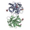
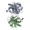
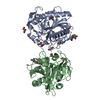
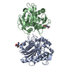
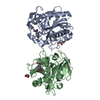
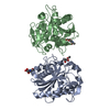
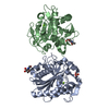
| Title | Crystal Structure of the complex of three phase partition treated lipase from Thermomyces lanuginosa with Lauric acid and P-nitrobenzaldehyde (PNB) at 2.1 resolution | ||||||
 Components Components | Lipase | ||||||
 Keywords Keywords | HYDROLASE | ||||||
| Function / homology |  Function and homology information Function and homology informationtriacylglycerol lipase / triacylglycerol lipase activity / lipid catabolic process Similarity search - Function | ||||||
| Biological species |   Thermomyces lanuginosus (fungus) Thermomyces lanuginosus (fungus) | ||||||
| Method |  X-RAY DIFFRACTION / SYNCHROTRON / MOLECULAR REPLACEMENT / Resolution: 2.1 Å X-RAY DIFFRACTION / SYNCHROTRON / MOLECULAR REPLACEMENT / Resolution: 2.1 Å | ||||||
 Authors Authors | Kumar, M. / Mukherjee, J. / Gupta, M.N. / Sinha, M. / Kaur, P. / Sharma, S. / Singh, T.P. | ||||||
 Citation Citation | Journal: TO BE PUBLISHED Title: Crystal Structure of the complex of three phase partition treated lipase from Thermomyces lanuginosa with Lauric acid and P-nitrobenzaldehyde (PNB) at 2.1 resolution Authors: Kumar, M. / Mukherjee, J. / Gupta, M.N. / Sinha, M. / Kaur, P. / Sharma, S. / Singh, T.P. | ||||||
| History |
|
- Structure visualization
| Structure viewer | Molecule: MolmilJmol/JSmol |
|---|
- Downloads & links
-Download
| PDBx/mmCIF format | 4kjx.cif.gz | 122.9 KB | Display | PDBx/mmCIF format |
|---|---|---|---|---|
| PDB format | pdb4kjx.ent.gz | 96.2 KB | Display | PDB format |
| PDBx/mmJSON format | 4kjx.json.gz | Tree view | PDBx/mmJSON format | |
| Others |  Other downloads Other downloads |
-Validation report
| Arichive directory | https://data.pdbj.org/pub/pdb/validation_reports/kj/4kjxftp://data.pdbj.org/pub/pdb/validation_reports/kj/4kjx | HTTPS FTP |
|---|
-Related structure data
| Related structure data | 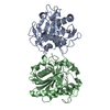 4ea6S S: Starting model for refinement |
|---|---|
| Similar structure data |


-Links
 PDBj
PDBj
- Assembly
| Deposited unit | 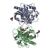
| ||||||||
|---|---|---|---|---|---|---|---|---|---|
| 1 |
| ||||||||
| Unit cell |
|
-Components
-Protein / Sugars , 2 types, 4 molecules AB
| #1: Protein | Mass: 29342.484 Da / Num. of mol.: 2 / Fragment: UNP residues 23-291 / Source method: isolated from a natural source / Source: (natural) Thermomyces lanuginosus (fungus) / References: UniProt: O59952, triacylglycerol lipase#2: Sugar |  Type: D-saccharide, beta linking / Mass: 221.208 Da / Num. of mol.: 2 Type: D-saccharide, beta linking / Mass: 221.208 Da / Num. of mol.: 2Source method: isolated from a genetically manipulated source Formula: C8H15NO6 |
|---|
-Non-polymers , 6 types, 280 molecules 


| #3: Chemical | ChemComp-GOL /  Mass: 92.094 Da / Num. of mol.: 6 / Source method: obtained synthetically / Formula: C3H8O3 Mass: 92.094 Da / Num. of mol.: 6 / Source method: obtained synthetically / Formula: C3H8O3#4: Chemical | ChemComp-ACT / |  Mass: 59.044 Da / Num. of mol.: 1 / Source method: obtained synthetically / Formula: C2H3O2 Mass: 59.044 Da / Num. of mol.: 1 / Source method: obtained synthetically / Formula: C2H3O2#5: Chemical |  Mass: 200.318 Da / Num. of mol.: 2 / Source method: obtained synthetically / Formula: C12H24O2 Mass: 200.318 Da / Num. of mol.: 2 / Source method: obtained synthetically / Formula: C12H24O2#6: Chemical | ChemComp-EDO / |  Mass: 62.068 Da / Num. of mol.: 1 / Source method: obtained synthetically / Formula: C2H6O2 Mass: 62.068 Da / Num. of mol.: 1 / Source method: obtained synthetically / Formula: C2H6O2#7: Chemical | ChemComp-XXH / |  Mass: 151.119 Da / Num. of mol.: 1 / Source method: obtained synthetically / Formula: C7H5NO3 Mass: 151.119 Da / Num. of mol.: 1 / Source method: obtained synthetically / Formula: C7H5NO3#8: Water | ChemComp-HOH / | Mass: 18.015 Da / Num. of mol.: 269 / Source method: isolated from a natural source / Formula: H2O |
|---|
-Details
| Has protein modification | Y |
|---|
-Experimental details
-Experiment
| Experiment | Method: X-RAY DIFFRACTION |
|---|
- Sample preparation
| Crystal | Density Matthews: 3.88 Å3/Da / Density % sol: 68.3 % |
|---|---|
| Crystal grow | Temperature: 298 K / Method: vapor diffusion, hanging drop / pH: 7.5 Details: 0.1M HEPES, O.1M Nacl, 1.6M Ammonium sulphate , pH 7.5, VAPOR DIFFUSION, HANGING DROP, temperature 298K |
-Data collection
| Diffraction | Mean temperature: 77 K |
|---|---|
| Diffraction source | Source: SYNCHROTRON / Site: ESRF  / Beamline: BM14 / Wavelength: 0.97 Å / Beamline: BM14 / Wavelength: 0.97 Å |
| Detector | Type: MARRESEARCH / Detector: CCD / Date: May 18, 2012 / Details: Mirror |
| Radiation | Monochromator: graphite / Protocol: SINGLE WAVELENGTH / Monochromatic (M) / Laue (L): M / Scattering type: x-ray |
| Radiation wavelength | Wavelength: 0.97 Å / Relative weight: 1 |
| Reflection | Resolution: 2.1→50 Å / Num. all: 98715 / Num. obs: 48911 / % possible obs: 98.6 % / Observed criterion σ(F): 0 / Observed criterion σ(I): 0 / Biso Wilson estimate: 40.5 Å2 / Rsym value: 0.086 / Net I/σ(I): 35.3 |
| Reflection shell | Resolution: 2.1→2.14 Å / Mean I/σ(I) obs: 2 / Num. unique all: 48911 / Rsym value: 0.443 / % possible all: 98.6 |
- Processing
| Software |
| ||||||||||||||||||||||||||||||||||||||||||||||||||||||||||||
|---|---|---|---|---|---|---|---|---|---|---|---|---|---|---|---|---|---|---|---|---|---|---|---|---|---|---|---|---|---|---|---|---|---|---|---|---|---|---|---|---|---|---|---|---|---|---|---|---|---|---|---|---|---|---|---|---|---|---|---|---|---|
| Refinement | Method to determine structure: MOLECULAR REPLACEMENT Starting model: 4EA6 Resolution: 2.1→45.83 Å / Cor.coef. Fo:Fc: 0.949 / Cor.coef. Fo:Fc free: 0.919 / SU B: 6.994 / SU ML: 0.156 / Cross valid method: THROUGHOUT / ESU R: 0.037 / ESU R Free: 0.037 / Stereochemistry target values: MAXIMUM LIKELIHOOD / Details: HYDROGENS HAVE BEEN ADDED IN THE RIDING POSITIONS
| ||||||||||||||||||||||||||||||||||||||||||||||||||||||||||||
| Solvent computation | Ion probe radii: 0.8 Å / Shrinkage radii: 0.8 Å / VDW probe radii: 1.2 Å / Solvent model: MASK | ||||||||||||||||||||||||||||||||||||||||||||||||||||||||||||
| Displacement parameters | Biso mean: 35.951 Å2
| ||||||||||||||||||||||||||||||||||||||||||||||||||||||||||||
| Refinement step | Cycle: LAST / Resolution: 2.1→45.83 Å
| ||||||||||||||||||||||||||||||||||||||||||||||||||||||||||||
| Refine LS restraints |
| ||||||||||||||||||||||||||||||||||||||||||||||||||||||||||||
| LS refinement shell | Resolution: 2.1→2.154 Å / Total num. of bins used: 20
|
