 Movie
Movie Controller


+ Open data

- Basic information
| Entry | Database: PDB / ID: 4kfz | ||||||
|---|---|---|---|---|---|---|---|
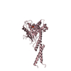
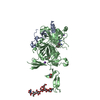
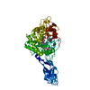
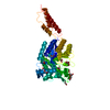
| Title | Crystal structure of LMO2 and anti-LMO2 VH complex | ||||||
 Components Components |
| ||||||
 Keywords Keywords | TRANSCRIPTION / ONCOPROTEIN / T-CELL LEUKEMIA / PROTO-ONCOGENE / DEVELOPMENTAL PROTEIN / LIM domain / transcription factor / nucleus | ||||||
| Function / homology |  Function and homology information Function and homology informationbHLH transcription factor binding / CD22 mediated BCR regulation / Fc epsilon receptor (FCERI) signaling / Classical antibody-mediated complement activation / Initial triggering of complement / immunoglobulin mediated immune response / FCGR activation / Role of LAT2/NTAL/LAB on calcium mobilization / immunoglobulin complex / Role of phospholipids in phagocytosis ...bHLH transcription factor binding / CD22 mediated BCR regulation / Fc epsilon receptor (FCERI) signaling / Classical antibody-mediated complement activation / Initial triggering of complement / immunoglobulin mediated immune response / FCGR activation / Role of LAT2/NTAL/LAB on calcium mobilization / immunoglobulin complex / Role of phospholipids in phagocytosis / Scavenging of heme from plasma / antigen binding / FCERI mediated Ca+2 mobilization / FCGR3A-mediated IL10 synthesis / Regulation of Complement cascade / Antigen activates B Cell Receptor (BCR) leading to generation of second messengers / Cell surface interactions at the vascular wall / transcription coregulator binding / FCGR3A-mediated phagocytosis / FCERI mediated MAPK activation / Regulation of actin dynamics for phagocytic cup formation / Immunoregulatory interactions between a Lymphoid and a non-Lymphoid cell / FCERI mediated NF-kB activation / RUNX1 regulates transcription of genes involved in differentiation of HSCs / transcription regulator complex / blood microparticle / DNA-binding transcription factor binding / Potential therapeutics for SARS / RNA polymerase II-specific DNA-binding transcription factor binding / transcription coactivator activity / immune response / positive regulation of transcription by RNA polymerase II / : / extracellular exosome / extracellular region / nucleoplasm / metal ion binding / identical protein binding / nucleus / plasma membrane Similarity search - Function | ||||||
| Biological species |  Homo sapiens (human) Homo sapiens (human) | ||||||
| Method |  X-RAY DIFFRACTION / SYNCHROTRON / MOLECULAR REPLACEMENT / Resolution: 2.8 Å X-RAY DIFFRACTION / SYNCHROTRON / MOLECULAR REPLACEMENT / Resolution: 2.8 Å | ||||||
 Authors Authors | Sewell, H. / Tanaka, T. / El Omari, K. / Cruz-Migoni, A. / Mancini, E.J. / Fuentes-Fernandez, N. / Chambers, J. / Rabbitts, T.H. | ||||||
 Citation Citation | Journal: Sci Rep / Year: 2014 Title: Conformational flexibility of the oncogenic protein LMO2 primes the formation of the multi-protein transcription complex. Authors: Sewell, H. / Tanaka, T. / Omari, K.E. / Mancini, E.J. / Cruz, A. / Fernandez-Fuentes, N. / Chambers, J. / Rabbitts, T.H. | ||||||
| History |
|
- Structure visualization
| Structure viewer | Molecule: MolmilJmol/JSmol |
|---|
- Downloads & links
-Download
| PDBx/mmCIF format | 4kfz.cif.gz | 228.8 KB | Display | PDBx/mmCIF format |
|---|---|---|---|---|
| PDB format | pdb4kfz.ent.gz | 186.3 KB | Display | PDB format |
| PDBx/mmJSON format | 4kfz.json.gz | Tree view | PDBx/mmJSON format | |
| Others |  Other downloads Other downloads |
-Validation report
| Arichive directory | https://data.pdbj.org/pub/pdb/validation_reports/kf/4kfzftp://data.pdbj.org/pub/pdb/validation_reports/kf/4kfz | HTTPS FTP |
|---|
-Related structure data
| Related structure data | 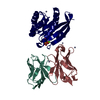 2uziS S: Starting model for refinement |
|---|---|
| Similar structure data |


-Links
 PDBj
PDBj


- Assembly
| Deposited unit | 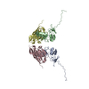
| ||||||||||||||||||||||||||||||||||||||||||||||||
|---|---|---|---|---|---|---|---|---|---|---|---|---|---|---|---|---|---|---|---|---|---|---|---|---|---|---|---|---|---|---|---|---|---|---|---|---|---|---|---|---|---|---|---|---|---|---|---|---|---|
| 1 | 
| ||||||||||||||||||||||||||||||||||||||||||||||||
| 2 | 
| ||||||||||||||||||||||||||||||||||||||||||||||||
| 3 | x 6
| ||||||||||||||||||||||||||||||||||||||||||||||||
| 4 | x 6
| ||||||||||||||||||||||||||||||||||||||||||||||||
| Unit cell |
| ||||||||||||||||||||||||||||||||||||||||||||||||
| Noncrystallographic symmetry (NCS) | NCS domain:
NCS domain segments:
NCS ensembles :
|
-Components
| #1: Protein | Mass: 17479.266 Da / Num. of mol.: 2 / Fragment: UNP residues 9-158 Source method: isolated from a genetically manipulated source Source: (gene. exp.) Homo sapiens (human) / Gene: LMO2, RBTN2, RBTNL1, RHOM2, TTG2 / Production host:  #2: Antibody | Mass: 14069.563 Da / Num. of mol.: 2 Source method: isolated from a genetically manipulated source Source: (gene. exp.) Homo sapiens (human) / Gene: Anti-LMO2 VH / Production host: #3: Chemical | ChemComp-ZN /   Mass: 65.409 Da / Num. of mol.: 8 / Source method: obtained synthetically / Formula: Zn Mass: 65.409 Da / Num. of mol.: 8 / Source method: obtained synthetically / Formula: Zn |
|---|
-Experimental details
-Experiment
| Experiment | Method: X-RAY DIFFRACTION |
|---|
- Sample preparation
| Crystal | Density Matthews: 2.88 Å3/Da / Density % sol: 57.24 % |
|---|---|
| Crystal grow | Temperature: 295 K / Method: vapor diffusion, sitting drop / pH: 6 Details: 100mM MES monohydrate pH 6.0, 0.8 M ammonium sulfate, and additive 1, 6 hexanediol, VAPOR DIFFUSION, SITTING DROP, temperature 295K |
-Data collection
| Diffraction | Mean temperature: 100 K |
|---|---|
| Diffraction source | Source: SYNCHROTRON / Site: Diamond  / Beamline: I02 / Wavelength: 0.979 Å / Beamline: I02 / Wavelength: 0.979 Å |
| Detector | Type: ADSC QUANTUM 315 / Detector: CCD / Date: Sep 19, 2009 |
| Radiation | Protocol: MAD / Monochromatic (M) / Laue (L): M / Scattering type: x-ray |
| Radiation wavelength | Wavelength: 0.979 Å / Relative weight: 1 |
| Reflection | Resolution: 2.8→50 Å / Num. all: 16868 / Num. obs: 16834 / % possible obs: 99.8 % / Redundancy: 2.9 % / Net I/σ(I): 10.8 |
| Reflection shell | Resolution: 2.8→2.95 Å / Redundancy: 3 % / Rmerge(I) obs: 0.73 / Mean I/σ(I) obs: 2.3 / % possible all: 100 |
- Processing
| Software |
| |||||||||||||||||||||||||||||||||||||||||||||||||||||||||||||||||||||||||||||||||||||||||||||||||||||||||||||||||||||||||||||
|---|---|---|---|---|---|---|---|---|---|---|---|---|---|---|---|---|---|---|---|---|---|---|---|---|---|---|---|---|---|---|---|---|---|---|---|---|---|---|---|---|---|---|---|---|---|---|---|---|---|---|---|---|---|---|---|---|---|---|---|---|---|---|---|---|---|---|---|---|---|---|---|---|---|---|---|---|---|---|---|---|---|---|---|---|---|---|---|---|---|---|---|---|---|---|---|---|---|---|---|---|---|---|---|---|---|---|---|---|---|---|---|---|---|---|---|---|---|---|---|---|---|---|---|---|---|---|
| Refinement | Method to determine structure: MOLECULAR REPLACEMENT Starting model: 2UZI (chain H) Resolution: 2.8→50 Å / Cor.coef. Fo:Fc: 0.918 / Cor.coef. Fo:Fc free: 0.909 / SU B: 32.498 / SU ML: 0.286 / Cross valid method: THROUGHOUT / ESU R Free: 0.374 / Stereochemistry target values: MAXIMUM LIKELIHOOD Details: HYDROGENS HAVE BEEN USED IF PRESENT IN THE INPUT, NON-CRYSTALLOGRAPHIC SYMMETRY (NCS) RESTRAINS WERE IMPOSED ON THE 2 VH#576/LMO2 DIMERS. TOWARDS THE END OF REFINEMENT, TLS ...Details: HYDROGENS HAVE BEEN USED IF PRESENT IN THE INPUT, NON-CRYSTALLOGRAPHIC SYMMETRY (NCS) RESTRAINS WERE IMPOSED ON THE 2 VH#576/LMO2 DIMERS. TOWARDS THE END OF REFINEMENT, TLS (TRANSLATION/LIBRATION/SCREW) VIBRATIONAL MOTION REFINEMENT WAS USED
| |||||||||||||||||||||||||||||||||||||||||||||||||||||||||||||||||||||||||||||||||||||||||||||||||||||||||||||||||||||||||||||
| Solvent computation | Ion probe radii: 0.8 Å / Shrinkage radii: 0.8 Å / VDW probe radii: 1.2 Å / Solvent model: MASK | |||||||||||||||||||||||||||||||||||||||||||||||||||||||||||||||||||||||||||||||||||||||||||||||||||||||||||||||||||||||||||||
| Displacement parameters | Biso mean: 75.405 Å2
| |||||||||||||||||||||||||||||||||||||||||||||||||||||||||||||||||||||||||||||||||||||||||||||||||||||||||||||||||||||||||||||
| Refinement step | Cycle: LAST / Resolution: 2.8→50 Å
| |||||||||||||||||||||||||||||||||||||||||||||||||||||||||||||||||||||||||||||||||||||||||||||||||||||||||||||||||||||||||||||
| Refine LS restraints |
| |||||||||||||||||||||||||||||||||||||||||||||||||||||||||||||||||||||||||||||||||||||||||||||||||||||||||||||||||||||||||||||
| Refine LS restraints NCS | Refine-ID: X-RAY DIFFRACTION / Type: interatomic distance / Weight position: 0.05
| |||||||||||||||||||||||||||||||||||||||||||||||||||||||||||||||||||||||||||||||||||||||||||||||||||||||||||||||||||||||||||||
| LS refinement shell | Resolution: 2.8→2.873 Å / Total num. of bins used: 20
| |||||||||||||||||||||||||||||||||||||||||||||||||||||||||||||||||||||||||||||||||||||||||||||||||||||||||||||||||||||||||||||
| Refinement TLS params. | Method: refined / Refine-ID: X-RAY DIFFRACTION
| |||||||||||||||||||||||||||||||||||||||||||||||||||||||||||||||||||||||||||||||||||||||||||||||||||||||||||||||||||||||||||||
| Refinement TLS group |
|
