 Movie
Movie Controller

[English] 日本語
 Yorodumi
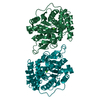
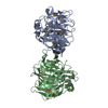
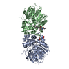
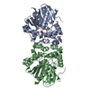
- PDB-4keq: Crystal structure of 4-pyridoxolactonase, 5-pyridoxolactone bound -
+ Open data

- Basic information
| Entry | Database: PDB / ID: 4keq | ||||||
|---|---|---|---|---|---|---|---|
| Title | Crystal structure of 4-pyridoxolactonase, 5-pyridoxolactone bound | ||||||
 Components Components | 4-pyridoxolactonase | ||||||
 Keywords Keywords | HYDROLASE / alpha-beta/beta-alpha fold / lactone | ||||||
| Function / homology |  Function and homology information Function and homology information4-pyridoxolactonase / 4-pyridoxolactonase activity / vitamin B6 catabolic process / metal ion binding Similarity search - Function | ||||||
| Biological species |  Mesorhizobium loti (bacteria) Mesorhizobium loti (bacteria) | ||||||
| Method |  X-RAY DIFFRACTION / SYNCHROTRON / MOLECULAR REPLACEMENT / Resolution: 2.279 Å X-RAY DIFFRACTION / SYNCHROTRON / MOLECULAR REPLACEMENT / Resolution: 2.279 Å | ||||||
 Authors Authors | Kobayashi, J. / Yoshikane, Y. / Baba, S. / Mizutani, K. / Takahashi, N. / Mikami, B. / Yagi, T. | ||||||
 Citation Citation | Journal: Acta Crystallogr.,Sect.F / Year: 2014 Title: Structure of 4-pyridoxolactonase from Mesorhizobium loti. Authors: Kobayashi, J. / Yoshikane, Y. / Yagi, T. / Baba, S. / Mizutani, K. / Takahashi, N. / Mikami, B. | ||||||
| History |
|
- Structure visualization
| Structure viewer | Molecule: MolmilJmol/JSmol |
|---|
- Downloads & links
-Download
| PDBx/mmCIF format | 4keq.cif.gz | 71 KB | Display | PDBx/mmCIF format |
|---|---|---|---|---|
| PDB format | pdb4keq.ent.gz | 51.1 KB | Display | PDB format |
| PDBx/mmJSON format | 4keq.json.gz | Tree view | PDBx/mmJSON format | |
| Others |  Other downloads Other downloads |
-Validation report
| Arichive directory | https://data.pdbj.org/pub/pdb/validation_reports/ke/4keqftp://data.pdbj.org/pub/pdb/validation_reports/ke/4keq | HTTPS FTP |
|---|
-Related structure data
| Related structure data | 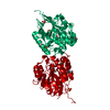 3aj3C  4kepC  3aj0 C: citing same article ( S: Starting model for refinement |
|---|---|
| Similar structure data |


-Links
 PDBj
PDBj

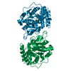
- Assembly
| Deposited unit | 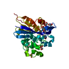
| ||||||||
|---|---|---|---|---|---|---|---|---|---|
| 1 | 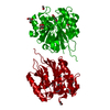
| ||||||||
| 2 | 
| ||||||||
| Unit cell |
|
-Components
-Protein , 1 types, 1 molecules A
| #1: Protein | Mass: 30725.635 Da / Num. of mol.: 1 Source method: isolated from a genetically manipulated source Source: (gene. exp.) Mesorhizobium loti (bacteria) / Strain: MAFF303099 / Gene: mlr6805 / Plasmid: pET21a / Production host: |
|---|
-Non-polymers , 5 types, 51 molecules 
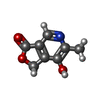


| #2: Chemical |  Mass: 65.409 Da / Num. of mol.: 2 / Source method: obtained synthetically / Formula: Zn Mass: 65.409 Da / Num. of mol.: 2 / Source method: obtained synthetically / Formula: Zn#3: Chemical |  Mass: 165.146 Da / Num. of mol.: 2 / Source method: obtained synthetically / Formula: C8H7NO3 Mass: 165.146 Da / Num. of mol.: 2 / Source method: obtained synthetically / Formula: C8H7NO3#4: Chemical |  Mass: 92.094 Da / Num. of mol.: 3 / Source method: obtained synthetically / Formula: C3H8O3 Mass: 92.094 Da / Num. of mol.: 3 / Source method: obtained synthetically / Formula: C3H8O3#5: Chemical |  Mass: 62.068 Da / Num. of mol.: 3 / Source method: obtained synthetically / Formula: C2H6O2 Mass: 62.068 Da / Num. of mol.: 3 / Source method: obtained synthetically / Formula: C2H6O2#6: Water | ChemComp-HOH / | Mass: 18.015 Da / Num. of mol.: 41 / Source method: isolated from a natural source / Formula: H2O |
|---|
-Experimental details
-Experiment
| Experiment | Method: X-RAY DIFFRACTION / Number of used crystals: 1 |
|---|
- Sample preparation
| Crystal | Density Matthews: 2.04 Å3/Da / Density % sol: 39.62 % |
|---|---|
| Crystal grow | Temperature: 277 K / Method: vapor diffusion, sitting drop Details: 20% PEG3350, 0.05M ammonium citrate, VAPOR DIFFUSION, SITTING DROP, temperature 277K |
-Data collection
| Diffraction | Mean temperature: 100 K |
|---|---|
| Diffraction source | Source: SYNCHROTRON / Site: SPring-8  / Beamline: BL44XU / Wavelength: 0.9 Å / Beamline: BL44XU / Wavelength: 0.9 Å |
| Detector | Detector: CCD / Date: May 25, 2012 |
| Radiation | Protocol: SINGLE WAVELENGTH / Monochromatic (M) / Laue (L): M / Scattering type: x-ray |
| Radiation wavelength | Wavelength: 0.9 Å / Relative weight: 1 |
| Reflection | Resolution: 2.279→50 Å / Num. obs: 11493 / % possible obs: 99.6 % / Redundancy: 4.2 % / Biso Wilson estimate: 26.71 Å2 / Rmerge(I) obs: 0.078 / Net I/σ(I): 30.3 |
| Reflection shell | Resolution: 2.28→2.32 Å / Redundancy: 3.8 % / Rmerge(I) obs: 0.228 / Mean I/σ(I) obs: 12.7 / % possible all: 98.8 |
- Processing
| Software |
| |||||||||||||||||||||||||||||||||||
|---|---|---|---|---|---|---|---|---|---|---|---|---|---|---|---|---|---|---|---|---|---|---|---|---|---|---|---|---|---|---|---|---|---|---|---|---|
| Refinement | Method to determine structure: MOLECULAR REPLACEMENT Starting model: 3AJ0 3aj0 Resolution: 2.279→36.182 Å / Occupancy max: 1 / Occupancy min: 0.19 / SU ML: 0.23 / σ(F): 1.34 / Phase error: 24.28 / Stereochemistry target values: ML
| |||||||||||||||||||||||||||||||||||
| Solvent computation | Shrinkage radii: 0.9 Å / VDW probe radii: 1.11 Å / Solvent model: FLAT BULK SOLVENT MODEL | |||||||||||||||||||||||||||||||||||
| Displacement parameters | Biso max: 60.59 Å2 / Biso mean: 23.3836 Å2 / Biso min: 8.06 Å2 | |||||||||||||||||||||||||||||||||||
| Refinement step | Cycle: LAST / Resolution: 2.279→36.182 Å
| |||||||||||||||||||||||||||||||||||
| Refine LS restraints |
| |||||||||||||||||||||||||||||||||||
| LS refinement shell | Refine-ID: X-RAY DIFFRACTION / Total num. of bins used: 4
|
