 Movie
Movie Controller

[English] 日本語
 Yorodumi
- PDB-4kdz: Crystal structure of tRNA/rRNA methyltransferase YibK from Escher... -
+ Open data

- Basic information
| Entry | Database: PDB / ID: 4kdz | ||||||
|---|---|---|---|---|---|---|---|
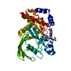
| Title | Crystal structure of tRNA/rRNA methyltransferase YibK from Escherichia coli (Target NYSGRC-012599) | ||||||
 Components Components | tRNA (cytidine(34)-2'-O)-methyltransferase | ||||||
 Keywords Keywords | TRANSFERASE / Structural Genomics / PSI-Biology / New York Structural Genomics Research Consortium / NYSGRC / SpoU methyltransferase / S-adenosyl-l-methionine | ||||||
| Function / homology |  Function and homology information Function and homology informationwobble position cytosine ribose methylation / wobble position uridine ribose methylation / tRNA (cytidine(34)-2'-O-ribose)-methyltransferase activity / tRNA (5-carboxymethylaminomethyluridine(34)-2'-O-ribose)-methyltransferase activity / tRNA (cytidine34-2'-O)-methyltransferase / RNA binding / cytoplasm Similarity search - Function | ||||||
| Biological species |  | ||||||
| Method |  X-RAY DIFFRACTION / SYNCHROTRON / MOLECULAR REPLACEMENT / Resolution: 2.32 Å X-RAY DIFFRACTION / SYNCHROTRON / MOLECULAR REPLACEMENT / Resolution: 2.32 Å | ||||||
 Authors Authors | Kim, J. / Toro, R. / Bhosle, R. / Almo, S.C. / New York Structural Genomics Research Consortium (NYSGRC) | ||||||
 Citation Citation | Journal: To be Published Title: Crystal structure of tRNA/rRNA methyltransferase YibK from Escherichia coli (Target NYSGRC-012599) Authors: Kim, J. / Toro, R. / Bhosle, R. / Almo, S.C. | ||||||
| History |
|
- Structure visualization
| Structure viewer | Molecule: MolmilJmol/JSmol |
|---|
- Downloads & links
-Download
| PDBx/mmCIF format | 4kdz.cif.gz | 44.8 KB | Display | PDBx/mmCIF format |
|---|---|---|---|---|
| PDB format | pdb4kdz.ent.gz | 31.1 KB | Display | PDB format |
| PDBx/mmJSON format | 4kdz.json.gz | Tree view | PDBx/mmJSON format | |
| Others |  Other downloads Other downloads |
-Validation report
| Arichive directory | https://data.pdbj.org/pub/pdb/validation_reports/kd/4kdzftp://data.pdbj.org/pub/pdb/validation_reports/kd/4kdz | HTTPS FTP |
|---|
-Related structure data
| Related structure data |  3n4jS S: Starting model for refinement |
|---|---|
| Similar structure data | |
| Other databases |

-Links
 PDBj
PDBj- Assembly
| Deposited unit | 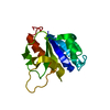
| ||||||||
|---|---|---|---|---|---|---|---|---|---|
| 1 | 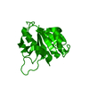
| ||||||||
| Unit cell |
|
-Components
| #1: Protein | Mass: 17747.225 Da / Num. of mol.: 1 Source method: isolated from a genetically manipulated source Source: (gene. exp.) References: UniProt: Q1R4Y9, tRNA (cytidine34-2'-O)-methyltransferase |
|---|---|
| #2: Water | ChemComp-HOH /  Mass: 18.015 Da / Num. of mol.: 58 / Source method: isolated from a natural source / Formula: H2O Mass: 18.015 Da / Num. of mol.: 58 / Source method: isolated from a natural source / Formula: H2O |
-Experimental details
-Experiment
| Experiment | Method: X-RAY DIFFRACTION / Number of used crystals: 1 |
|---|
- Sample preparation
| Crystal | Density Matthews: 4.56 Å3/Da / Density % sol: 73.03 % |
|---|---|
| Crystal grow | Temperature: 293 K / Method: vapor diffusion, sitting drop / pH: 7.5 Details: 0.1M HEPES (pH7.5) 1.4M sodium citrate tribasic dihydrate, VAPOR DIFFUSION, SITTING DROP, temperature 293K |
-Data collection
| Diffraction | Mean temperature: 100 K |
|---|---|
| Diffraction source | Source: SYNCHROTRON / Site: NSLS  / Beamline: X29A / Wavelength: 1.075 / Beamline: X29A / Wavelength: 1.075 |
| Detector | Type: ADSC QUANTUM 315r / Detector: CCD / Date: Jun 10, 2011 |
| Radiation | Monochromator: Double silicon(111) crystal / Protocol: SINGLE WAVELENGTH / Monochromatic (M) / Laue (L): M / Scattering type: x-ray |
| Radiation wavelength | Wavelength: 1.075 Å / Relative weight: 1 |
| Reflection | Resolution: 2.32→50 Å / Num. all: 14445 / Num. obs: 14428 / % possible obs: 99.9 % / Redundancy: 9 % / Rmerge(I) obs: 0.113 / Net I/σ(I): 17.8 |
| Reflection shell | Resolution: 2.32→2.36 Å / Redundancy: 9.2 % / Rmerge(I) obs: 0.93 / Mean I/σ(I) obs: 2.7 / % possible all: 100 |
- Processing
| Software |
| ||||||||||||||||||||||||||||||||||||||||||||||||||||||||||||||||||||||||||||||||||||||||||||||||||||||||||||||||||||||||||||||||||||||||||||||||||||||||||||||||||||||||||
|---|---|---|---|---|---|---|---|---|---|---|---|---|---|---|---|---|---|---|---|---|---|---|---|---|---|---|---|---|---|---|---|---|---|---|---|---|---|---|---|---|---|---|---|---|---|---|---|---|---|---|---|---|---|---|---|---|---|---|---|---|---|---|---|---|---|---|---|---|---|---|---|---|---|---|---|---|---|---|---|---|---|---|---|---|---|---|---|---|---|---|---|---|---|---|---|---|---|---|---|---|---|---|---|---|---|---|---|---|---|---|---|---|---|---|---|---|---|---|---|---|---|---|---|---|---|---|---|---|---|---|---|---|---|---|---|---|---|---|---|---|---|---|---|---|---|---|---|---|---|---|---|---|---|---|---|---|---|---|---|---|---|---|---|---|---|---|---|---|---|---|---|
| Refinement | Method to determine structure: MOLECULAR REPLACEMENT Starting model: PDB ENTRY 3N4J Resolution: 2.32→42.25 Å / Cor.coef. Fo:Fc: 0.947 / Cor.coef. Fo:Fc free: 0.928 / SU B: 4.993 / SU ML: 0.12 / Cross valid method: THROUGHOUT / ESU R: 0.181 / ESU R Free: 0.171 / Stereochemistry target values: MAXIMUM LIKELIHOOD / Details: HYDROGENS HAVE BEEN USED IF PRESENT IN THE INPUT
| ||||||||||||||||||||||||||||||||||||||||||||||||||||||||||||||||||||||||||||||||||||||||||||||||||||||||||||||||||||||||||||||||||||||||||||||||||||||||||||||||||||||||||
| Solvent computation | Ion probe radii: 0.8 Å / Shrinkage radii: 0.8 Å / VDW probe radii: 1.2 Å / Solvent model: MASK | ||||||||||||||||||||||||||||||||||||||||||||||||||||||||||||||||||||||||||||||||||||||||||||||||||||||||||||||||||||||||||||||||||||||||||||||||||||||||||||||||||||||||||
| Displacement parameters | Biso mean: 29.503 Å2
| ||||||||||||||||||||||||||||||||||||||||||||||||||||||||||||||||||||||||||||||||||||||||||||||||||||||||||||||||||||||||||||||||||||||||||||||||||||||||||||||||||||||||||
| Refinement step | Cycle: LAST / Resolution: 2.32→42.25 Å
| ||||||||||||||||||||||||||||||||||||||||||||||||||||||||||||||||||||||||||||||||||||||||||||||||||||||||||||||||||||||||||||||||||||||||||||||||||||||||||||||||||||||||||
| Refine LS restraints |
| ||||||||||||||||||||||||||||||||||||||||||||||||||||||||||||||||||||||||||||||||||||||||||||||||||||||||||||||||||||||||||||||||||||||||||||||||||||||||||||||||||||||||||
| LS refinement shell | Resolution: 2.319→2.379 Å / Total num. of bins used: 20
|
