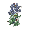
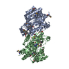
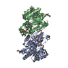
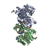
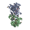
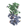
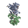
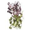
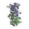
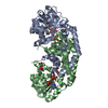
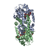
- PDB-4k8k: Crystal structure of probable sugar kinase protein from Rhizobium... -
+
Open data
ID or keywords:
Loading...
-
Basic information
Entry
Database: PDB / ID: 4k8k
Title
Crystal structure of probable sugar kinase protein from Rhizobium etli CFN 42 complexed with 1-(4-methoxyphenyl)-1-cyclopropane and 2-aminoperimidine
Components
sugar kinase
Keywords
TRANSFERASE / STRUCTURAL GENOMICS / PROTEIN STRUCTURE INITIATIVE / NEW YORK STRUCTURAL GENOMIX RESEARCHCONSORTIUM / NYSGRC / PSI-Biology / New York Structural Genomics Research Consortium
Resolution: 1.49→50 Å / Num. obs: 111895 / % possible obs: 99.7 % / Redundancy: 7.1 % / Rmerge(I) obs: 0.083 / Χ2: 0.979 / Net I/σ(I): 8.2
Reflection shell
Resolution (Å)
Redundancy (%)
Rmerge(I) obs
Num. unique all
Χ2
Diffraction-ID
% possible all
1.49-1.52
6.8
0.594
5519
0.824
1
99.1
1.52-1.54
6.9
0.513
5480
0.878
1
99.5
1.54-1.57
7
0.435
5486
0.92
1
99.2
1.57-1.61
7
0.384
5558
0.968
1
99.6
1.61-1.64
7
0.349
5523
1.034
1
99.5
1.64-1.68
7
0.307
5545
0.921
1
99.7
1.68-1.72
7
0.283
5544
0.987
1
99.7
1.72-1.77
7
0.228
5532
0.947
1
99.6
1.77-1.82
7
0.194
5563
0.964
1
99.6
1.82-1.88
7.1
0.181
5554
0.991
1
99.7
1.88-1.94
7.1
0.145
5576
0.99
1
99.7
1.94-2.02
7.1
0.126
5589
1.011
1
99.7
2.02-2.11
7.2
0.108
5578
1.009
1
99.9
2.11-2.23
7.2
0.099
5608
1.011
1
100
2.23-2.37
7.3
0.089
5615
1.013
1
100
2.37-2.55
7.3
0.081
5616
1.077
1
100
2.55-2.8
7.2
0.075
5663
1.077
1
100
2.8-3.21
7.2
0.064
5685
1.013
1
100
3.21-4.04
7
0.056
5731
0.937
1
99.7
4.04-50
6.9
0.053
5930
0.969
1
99.4
-
Phasing
Phasing
Method: molecular replacement
-
Processing
Software
Name
Version
Classification
NB
SCALEPACK
datascaling
PHASER
phasing
REFMAC
refinement
PDB_EXTRACT
3.11
dataextraction
CBASS
datacollection
HKL-2000
datareduction
Refinement
Method to determine structure: MOLECULAR REPLACEMENT / Resolution: 1.5→39.65 Å / Cor.coef. Fo:Fc: 0.967 / Cor.coef. Fo:Fc free: 0.957 / WRfactor Rfree: 0.2324 / WRfactor Rwork: 0.1871 / Occupancy max: 1 / Occupancy min: 0.4 / FOM work R set: 0.8097 / SU B: 1.815 / SU ML: 0.033 / SU R Cruickshank DPI: 0.0203 / SU Rfree: 0.017 / Cross valid method: THROUGHOUT / σ(F): 0 / ESU R: 0.02 / ESU R Free: 0.017 / Stereochemistry target values: MAXIMUM LIKELIHOOD Details: THE RESIDUAL FLAT DENSITY BETWEEN ILE-307 AND SER-245 IS DUE TO THE TRACE AMOUNTS OF EXOGENOUS ADP OR ITS ANALOGS. HYDROGENS HAVE BEEN USED IF PRESENT IN THE INPUT U VALUES : RESIDUAL ONLY
Rfactor
Num. reflection
% reflection
Selection details
Rfree
0.2185
5501
5 %
RANDOM
Rwork
0.1806
-
-
-
obs
0.1825
109969
99.69 %
-
Solvent computation
Ion probe radii: 0.8 Å / Shrinkage radii: 0.8 Å / VDW probe radii: 1.2 Å / Solvent model: MASK
In the structure databanks used in Yorodumi, some data are registered as the other names, "COVID-19 virus" and "2019-nCoV". Here are the details of the virus and the list of structure data.
Jan 31, 2019. EMDB accession codes are about to change! (news from PDBe EMDB page)
EMDB accession codes are about to change! (news from PDBe EMDB page)
The allocation of 4 digits for EMDB accession codes will soon come to an end. Whilst these codes will remain in use, new EMDB accession codes will include an additional digit and will expand incrementally as the available range of codes is exhausted. The current 4-digit format prefixed with “EMD-” (i.e. EMD-XXXX) will advance to a 5-digit format (i.e. EMD-XXXXX), and so on. It is currently estimated that the 4-digit codes will be depleted around Spring 2019, at which point the 5-digit format will come into force.
The EM Navigator/Yorodumi systems omit the EMD- prefix.
Related info.:Q: What is EMD? / ID/Accession-code notation in Yorodumi/EM Navigator
Yorodumi is a browser for structure data from EMDB, PDB, SASBDB, etc.
This page is also the successor to EM Navigator detail page, and also detail information page/front-end page for Omokage search.
The word "yorodu" (or yorozu) is an old Japanese word meaning "ten thousand". "mi" (miru) is to see.
Related info.:EMDB / PDB / SASBDB / Comparison of 3 databanks / Yorodumi Search / Aug 31, 2016. New EM Navigator & Yorodumi / Yorodumi Papers / Jmol/JSmol / Function and homology information / Changes in new EM Navigator and Yorodumi
 Movie
Movie Controller

 Yorodumi
 Open data
 Basic information
 Components
 Keywords
 Function and homology information
 Rhizobium etli (bacteria)
 X-RAY DIFFRACTION /
  Authors
 Citation
 Structure visualization
 Downloads & links
 Other downloads
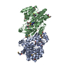


 PDBj
PDBj
 Assembly
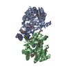


 Mass: 267.241 Da / Num. of mol.: 2 / Source method: obtained synthetically / Formula: C10H13N5O4
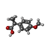
 Mass: 192.211 Da / Num. of mol.: 2 / Source method: obtained synthetically / Formula: C11H12O3
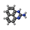
 Mass: 183.209 Da / Num. of mol.: 8 / Source method: obtained synthetically / Formula: C11H9N3
 Mass: 39.098 Da / Num. of mol.: 2 / Source method: obtained synthetically / Formula: K
 Sample preparation
 / Beamline: X29A / Wavelength: 1.075 Å
 Processing