 Movie
Movie Controller


+ Open data

- Basic information
| Entry | Database: PDB / ID: 4k7e | ||||||
|---|---|---|---|---|---|---|---|
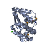
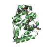
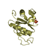
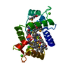
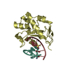
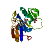
| Title | Crystal structure of Junin virus nucleoprotein | ||||||
 Components Components | Nucleoprotein | ||||||
 Keywords Keywords | VIRAL PROTEIN / alpha/beta/alpha sandwich / nucleoprotein | ||||||
| Function / homology |  Function and homology information Function and homology informationsymbiont-mediated suppression of host cytoplasmic pattern recognition receptor signaling pathway via inhibition of IKBKE activity / RNA-templated viral transcription / negative stranded viral RNA replication / helical viral capsid / viral nucleocapsid / Hydrolases; Acting on ester bonds; Exoribonucleases producing 5'-phosphomonoesters / host cell cytoplasm / ribonucleoprotein complex / hydrolase activity / RNA binding / metal ion binding Similarity search - Function | ||||||
| Biological species |  Junin virus Junin virus | ||||||
| Method |  X-RAY DIFFRACTION / SYNCHROTRON / MOLECULAR REPLACEMENT / Resolution: 2.2 Å X-RAY DIFFRACTION / SYNCHROTRON / MOLECULAR REPLACEMENT / Resolution: 2.2 Å | ||||||
 Authors Authors | Zhang, Y.J. / Li, L. / Liu, X. / Dong, S.S. / Wang, W.M. / Huo, T. / Rao, Z.H. / Yang, C. | ||||||
 Citation Citation | Journal: J.Gen.Virol. / Year: 2013 Title: Crystal structure of Junin virus nucleoprotein Authors: Zhang, Y.J. / Li, L. / Liu, X. / Dong, S.S. / Wang, W.M. / Huo, T. / Guo, Y. / Rao, Z.H. / Yang, C. | ||||||
| History |
|
- Structure visualization
| Structure viewer | Molecule: MolmilJmol/JSmol |
|---|
- Downloads & links
-Download
| PDBx/mmCIF format | 4k7e.cif.gz | 46 KB | Display | PDBx/mmCIF format |
|---|---|---|---|---|
| PDB format | pdb4k7e.ent.gz | 30.8 KB | Display | PDB format |
| PDBx/mmJSON format | 4k7e.json.gz | Tree view | PDBx/mmJSON format | |
| Others |  Other downloads Other downloads |
-Validation report
| Arichive directory | https://data.pdbj.org/pub/pdb/validation_reports/k7/4k7eftp://data.pdbj.org/pub/pdb/validation_reports/k7/4k7e | HTTPS FTP |
|---|
-Related structure data
| Similar structure data |
|---|


-Links
 PDBj
PDBj- Assembly
| Deposited unit | 
| ||||||||
|---|---|---|---|---|---|---|---|---|---|
| 1 |
| ||||||||
| 2 | 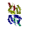
| ||||||||
| Unit cell |
|
-Components
| #1: Protein | Mass: 26667.738 Da / Num. of mol.: 1 / Fragment: C-TERMINAL DOMAIN, residues 341-564 Source method: isolated from a genetically manipulated source Source: (gene. exp.) Junin virus / Gene: N / Plasmid: pGEX-6p-1 / Production host:  |
|---|---|
| #2: Water | ChemComp-HOH /  Mass: 18.015 Da / Num. of mol.: 78 / Source method: isolated from a natural source / Formula: H2O Mass: 18.015 Da / Num. of mol.: 78 / Source method: isolated from a natural source / Formula: H2O |
| Has protein modification | Y |
-Experimental details
-Experiment
| Experiment | Method: X-RAY DIFFRACTION / Number of used crystals: 1 |
|---|
- Sample preparation
| Crystal | Density Matthews: 2.02 Å3/Da / Density % sol: 39.16 % |
|---|---|
| Crystal grow | Temperature: 289 K / Method: vapor diffusion, hanging drop / pH: 8 Details: 0.1M Tris, 2.2M ammonium sulfate, pH 8.0, VAPOR DIFFUSION, HANGING DROP, temperature 289K |
-Data collection
| Diffraction | Mean temperature: 100 K |
|---|---|
| Diffraction source | Source: SYNCHROTRON / Site: Photon Factory  / Beamline: BL-17A / Wavelength: 1 Å / Beamline: BL-17A / Wavelength: 1 Å |
| Detector | Type: ADSC QUANTUM 270 / Detector: CCD / Date: Dec 18, 2012 |
| Radiation | Protocol: SINGLE WAVELENGTH / Monochromatic (M) / Laue (L): M / Scattering type: x-ray |
| Radiation wavelength | Wavelength: 1 Å / Relative weight: 1 |
| Reflection | Resolution: 2.2→39.26 Å / Num. all: 11382 / Num. obs: 11370 / % possible obs: 99.8 % / Observed criterion σ(F): 3 / Observed criterion σ(I): 3 / Biso Wilson estimate: 32.11 Å2 |
| Reflection shell | Resolution: 2.2→2.24 Å / Redundancy: 10.7 % / Rmerge(I) obs: 0.083 / Mean I/σ(I) obs: 53.5 / Num. unique all: 11382 / % possible all: 99.8 |
- Processing
| Software |
| |||||||||||||||||||||||||||||||||||
|---|---|---|---|---|---|---|---|---|---|---|---|---|---|---|---|---|---|---|---|---|---|---|---|---|---|---|---|---|---|---|---|---|---|---|---|---|
| Refinement | Method to determine structure: MOLECULAR REPLACEMENT / Resolution: 2.2→39.256 Å / Occupancy max: 1 / Occupancy min: 0.39 / FOM work R set: 0.8133 / SU ML: 0.23 / σ(F): 0 / Phase error: 24.22 / Stereochemistry target values: ML
| |||||||||||||||||||||||||||||||||||
| Solvent computation | Shrinkage radii: 0.83 Å / VDW probe radii: 1.1 Å / Solvent model: FLAT BULK SOLVENT MODEL / Bsol: 45.851 Å2 / ksol: 0.373 e/Å3 | |||||||||||||||||||||||||||||||||||
| Displacement parameters | Biso max: 88.72 Å2 / Biso mean: 41.8034 Å2 / Biso min: 20 Å2
| |||||||||||||||||||||||||||||||||||
| Refinement step | Cycle: LAST / Resolution: 2.2→39.256 Å
| |||||||||||||||||||||||||||||||||||
| Refine LS restraints |
| |||||||||||||||||||||||||||||||||||
| LS refinement shell | Refine-ID: X-RAY DIFFRACTION / Total num. of bins used: 4
|
