 Movie
Movie Controller

[English] 日本語
 Yorodumi
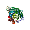
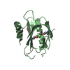
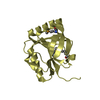
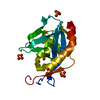
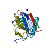
- PDB-4k0c: Crystal Structure of the computationally designed serine hydrolas... -
+ Open data

- Basic information
| Entry | Database: PDB / ID: 4k0c | ||||||
|---|---|---|---|---|---|---|---|
| Title | Crystal Structure of the computationally designed serine hydrolase. Northeast Structural Genomics Consortium (NESG) Target OR317 | ||||||
 Components Components | designed serine hydrolase | ||||||
 Keywords Keywords | Structural Genomics / Unknown Function / PSI-Biology / Protein Structure Initiative / Northeast Structural Genomics Consortium / NESG | ||||||
| Function / homology | Leucine Aminopeptidase, subunit E, domain 1 / Leucine Aminopeptidase, subunit E; domain 1 / 3-Layer(aba) Sandwich / Alpha Beta Function and homology information Function and homology information | ||||||
| Biological species | synthetic construct (others) | ||||||
| Method |  X-RAY DIFFRACTION / SYNCHROTRON / MOLECULAR REPLACEMENT / Resolution: 3.002 Å X-RAY DIFFRACTION / SYNCHROTRON / MOLECULAR REPLACEMENT / Resolution: 3.002 Å | ||||||
 Authors Authors | Kuzin, A. / Lew, S. / Rajagopalan, S. / Seetharaman, J. / Maglaqui, M. / Xiao, R. / Lee, D. / Everett, J.K. / Acton, T.B. / Baker, D. ...Kuzin, A. / Lew, S. / Rajagopalan, S. / Seetharaman, J. / Maglaqui, M. / Xiao, R. / Lee, D. / Everett, J.K. / Acton, T.B. / Baker, D. / Montelione, G.T. / Tong, L. / Hunt, J.F. / Northeast Structural Genomics Consortium (NESG) | ||||||
 Citation Citation | Journal: To be Published Title: Northeast Structural Genomics Consortium Target OR317 Authors: Kuzin, A. / Lew, S. / Rajagopalan, S. / Seetharaman, J. / Maglaqui, M. / Xiao, R. / Lee, D. / Everett, J.K. / Acton, T.B. / Baker, D. / Montelione, G.T. / Tong, L. / Hunt, J.F. | ||||||
| History |
|
- Structure visualization
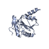
| Structure viewer | Molecule: MolmilJmol/JSmol |
|---|
- Downloads & links
-Download
| PDBx/mmCIF format | 4k0c.cif.gz | 69.8 KB | Display | PDBx/mmCIF format |
|---|---|---|---|---|
| PDB format | pdb4k0c.ent.gz | 52.2 KB | Display | PDB format |
| PDBx/mmJSON format | 4k0c.json.gz | Tree view | PDBx/mmJSON format | |
| Others |  Other downloads Other downloads |
-Validation report
| Arichive directory | https://data.pdbj.org/pub/pdb/validation_reports/k0/4k0cftp://data.pdbj.org/pub/pdb/validation_reports/k0/4k0c | HTTPS FTP |
|---|
-Related structure data
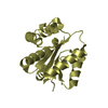
| Related structure data | 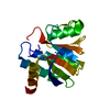 4essS S: Starting model for refinement |
|---|---|
| Similar structure data | |
| Other databases |


-Links
 PDBj
PDBj- Assembly
| Deposited unit | 
| ||||||||
|---|---|---|---|---|---|---|---|---|---|
| 1 | 
| ||||||||
| 2 | 
| ||||||||
| Unit cell |
|
-Components
| #1: Protein | Mass: 17873.129 Da / Num. of mol.: 2 Source method: isolated from a genetically manipulated source Source: (gene. exp.) synthetic construct (others) Has protein modification | Y | |
|---|
-Experimental details
-Experiment
| Experiment | Method: X-RAY DIFFRACTION / Number of used crystals: 1 |
|---|
- Sample preparation
| Crystal | Density Matthews: 2.05 Å3/Da / Density % sol: 39.98 % |
|---|---|
| Crystal grow | Temperature: 277 K / Method: microbatch under oil / pH: 6 Details: Protein solution: 100mM NaCl, 5mM DTT, 0.02% NaN3, 10mM Tris-HCl (pH 7.5) . Reservoir solution:0.1m magnesium sulfate heptahydrate, 0.1 MES, PEG 400 40% v/v., microbatch under oil, temperature 277K |
-Data collection
| Diffraction | Mean temperature: 100 K |
|---|---|
| Diffraction source | Source: SYNCHROTRON / Site: NSLS  / Beamline: X4A / Wavelength: 0.979 Å / Beamline: X4A / Wavelength: 0.979 Å |
| Detector | Type: ADSC QUANTUM 4 / Detector: CCD / Date: Jan 22, 2013 / Details: mirrors |
| Radiation | Monochromator: Si 111 CHANNEL / Protocol: SINGLE WAVELENGTH / Monochromatic (M) / Laue (L): M / Scattering type: x-ray |
| Radiation wavelength | Wavelength: 0.979 Å / Relative weight: 1 |
| Reflection | Resolution: 3→50 Å / Num. obs: 10205 / % possible obs: 90 % / Observed criterion σ(I): -3 / Redundancy: 5.4 % / Biso Wilson estimate: 48.52 Å2 / Rmerge(I) obs: 0.114 / Net I/σ(I): 9.7 |
- Processing
| Software |
| |||||||||||||||||||||||||||||||||||
|---|---|---|---|---|---|---|---|---|---|---|---|---|---|---|---|---|---|---|---|---|---|---|---|---|---|---|---|---|---|---|---|---|---|---|---|---|
| Refinement | Method to determine structure: MOLECULAR REPLACEMENT Starting model: PDB ENTRY 4ESS Resolution: 3.002→37.287 Å / Occupancy max: 1 / Occupancy min: 0.77 / FOM work R set: 0.816 / SU ML: 0.37 / Cross valid method: THROUGHOUT / σ(F): 1.45 / Phase error: 25.12 / Stereochemistry target values: ML
| |||||||||||||||||||||||||||||||||||
| Solvent computation | Shrinkage radii: 0.9 Å / VDW probe radii: 1.11 Å / Solvent model: FLAT BULK SOLVENT MODEL | |||||||||||||||||||||||||||||||||||
| Displacement parameters | Biso max: 97.86 Å2 / Biso mean: 34.838 Å2 / Biso min: 9.25 Å2 | |||||||||||||||||||||||||||||||||||
| Refinement step | Cycle: LAST / Resolution: 3.002→37.287 Å
| |||||||||||||||||||||||||||||||||||
| Refine LS restraints |
| |||||||||||||||||||||||||||||||||||
| LS refinement shell | Refine-ID: X-RAY DIFFRACTION / Total num. of bins used: 4
|
