 Movie
Movie Controller

[English] 日本語
 Yorodumi
- PDB-4jza: Crystal structure of a Legionella phosphoinositide phosphatase: i... -
+ Open data

- Basic information
| Entry | Database: PDB / ID: 4jza | ||||||
|---|---|---|---|---|---|---|---|
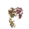
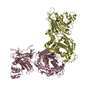
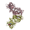
| Title | Crystal structure of a Legionella phosphoinositide phosphatase: insights into lipid metabolism in pathogen host interaction | ||||||
 Components Components | Uncharacterized protein | ||||||
 Keywords Keywords | HYDROLASE / alpha beta fold / phosphatase | ||||||
| Function / homology |  Function and homology information Function and homology informationPeroxidase; domain 1 - #60 / Four Helix Bundle (Hemerythrin (Met), subunit A) - #1720 / Phosphoinositide phosphatase insertion domain / Phosphoinositide phosphatase, C-terminal / Phosphoinositide phosphatase insertion domain / Phosphoinositide phosphatase C-terminal domain / Peroxidase; domain 1 / Four Helix Bundle (Hemerythrin (Met), subunit A) / Up-down Bundle / Orthogonal Bundle / Mainly Alpha Similarity search - Domain/homology | ||||||
| Biological species |  Legionella longbeachae NSW150 (bacteria) Legionella longbeachae NSW150 (bacteria) | ||||||
| Method |  X-RAY DIFFRACTION / SYNCHROTRON / SAD / Resolution: 2.584 Å X-RAY DIFFRACTION / SYNCHROTRON / SAD / Resolution: 2.584 Å | ||||||
 Authors Authors | Toulabi, L. / Wu, X. / Cheng, Y. / Mao, Y. | ||||||
 Citation Citation | Journal: J.Biol.Chem. / Year: 2013 Title: Identification and structural characterization of a legionella phosphoinositide phosphatase. Authors: Toulabi, L. / Wu, X. / Cheng, Y. / Mao, Y. | ||||||
| History |
|
- Structure visualization
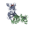
| Structure viewer | Molecule: MolmilJmol/JSmol |
|---|
- Downloads & links
-Download
| PDBx/mmCIF format | 4jza.cif.gz | 643 KB | Display | PDBx/mmCIF format |
|---|---|---|---|---|
| PDB format | pdb4jza.ent.gz | 539 KB | Display | PDB format |
| PDBx/mmJSON format | 4jza.json.gz | Tree view | PDBx/mmJSON format | |
| Others |  Other downloads Other downloads |
-Validation report
| Arichive directory | https://data.pdbj.org/pub/pdb/validation_reports/jz/4jzaftp://data.pdbj.org/pub/pdb/validation_reports/jz/4jza | HTTPS FTP |
|---|
-Related structure data
| Related structure data | |
|---|---|
| Similar structure data |


-Links
 PDBj
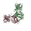
PDBj- Assembly
| Deposited unit | 
| ||||||||||||
|---|---|---|---|---|---|---|---|---|---|---|---|---|---|
| 1 | 
| ||||||||||||
| 2 | 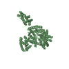
| ||||||||||||
| 3 |
| ||||||||||||
| Unit cell |
| ||||||||||||
| Noncrystallographic symmetry (NCS) | NCS oper:
|
-Components
| #1: Protein | Mass: 95049.992 Da / Num. of mol.: 2 / Source method: isolated from a natural source / Source: (natural) Legionella longbeachae NSW150 (bacteria) / Strain: NSW150References: UniProt: D3HMN4, phosphatidylinositol-3-phosphatase #2: Water | ChemComp-HOH / |  Mass: 18.015 Da / Num. of mol.: 161 / Source method: isolated from a natural source / Formula: H2O Mass: 18.015 Da / Num. of mol.: 161 / Source method: isolated from a natural source / Formula: H2OHas protein modification | Y | |
|---|
-Experimental details
-Experiment
| Experiment | Method: X-RAY DIFFRACTION / Number of used crystals: 1 |
|---|
- Sample preparation
| Crystal | Density Matthews: 3.7 Å3/Da / Density % sol: 66.75 % |
|---|---|
| Crystal grow | Temperature: 277 K / Method: vapor diffusion, hanging drop / pH: 7 Details: 0.1M HEPES (pH 7.0), 0.1M succinic acid, and 6.5% (wt/vol) PEG3350, VAPOR DIFFUSION, HANGING DROP, temperature 277K |
-Data collection
| Diffraction | Mean temperature: 100 K |
|---|---|
| Diffraction source | Source: SYNCHROTRON / Site: CHESS  / Beamline: A1 / Wavelength: 0.978 Å / Beamline: A1 / Wavelength: 0.978 Å |
| Detector | Type: ADSC QUANTUM 210 / Detector: CCD / Date: Jan 1, 2012 / Details: mirrors |
| Radiation | Monochromator: GRAPHITE / Protocol: SINGLE WAVELENGTH / Monochromatic (M) / Laue (L): M / Scattering type: x-ray |
| Radiation wavelength | Wavelength: 0.978 Å / Relative weight: 1 |
| Reflection | Resolution: 2.58→130.93 Å / Num. all: 167313 / Num. obs: 164803 / % possible obs: 98.5 % / Observed criterion σ(F): 1 / Observed criterion σ(I): 1 / Rmerge(I) obs: 0.099 / Net I/σ(I): 10.96 |
- Processing
| Software |
| ||||||||||||||||||||||||||||||||||||||||||||||||||||||||||||||||||||||||||||||||||||||||||||||||||||||||||||||||||||||||||||||||||||||||||||||||||||||||||||||||||||||||||||||||||||||
|---|---|---|---|---|---|---|---|---|---|---|---|---|---|---|---|---|---|---|---|---|---|---|---|---|---|---|---|---|---|---|---|---|---|---|---|---|---|---|---|---|---|---|---|---|---|---|---|---|---|---|---|---|---|---|---|---|---|---|---|---|---|---|---|---|---|---|---|---|---|---|---|---|---|---|---|---|---|---|---|---|---|---|---|---|---|---|---|---|---|---|---|---|---|---|---|---|---|---|---|---|---|---|---|---|---|---|---|---|---|---|---|---|---|---|---|---|---|---|---|---|---|---|---|---|---|---|---|---|---|---|---|---|---|---|---|---|---|---|---|---|---|---|---|---|---|---|---|---|---|---|---|---|---|---|---|---|---|---|---|---|---|---|---|---|---|---|---|---|---|---|---|---|---|---|---|---|---|---|---|---|---|---|---|
| Refinement | Method to determine structure: SAD / Resolution: 2.584→49.44 Å / Cor.coef. Fo:Fc: 0.949 / Cor.coef. Fo:Fc free: 0.923 / SU B: 19.411 / SU ML: 0.189 / Cross valid method: THROUGHOUT / ESU R: 0.372 / ESU R Free: 0.258 / Stereochemistry target values: MAXIMUM LIKELIHOOD / Details: HYDROGENS HAVE BEEN ADDED IN THE RIDING POSITIONS
| ||||||||||||||||||||||||||||||||||||||||||||||||||||||||||||||||||||||||||||||||||||||||||||||||||||||||||||||||||||||||||||||||||||||||||||||||||||||||||||||||||||||||||||||||||||||
| Solvent computation | Ion probe radii: 0.8 Å / Shrinkage radii: 0.8 Å / VDW probe radii: 1.2 Å / Solvent model: MASK | ||||||||||||||||||||||||||||||||||||||||||||||||||||||||||||||||||||||||||||||||||||||||||||||||||||||||||||||||||||||||||||||||||||||||||||||||||||||||||||||||||||||||||||||||||||||
| Displacement parameters | Biso mean: 58.829 Å2
| ||||||||||||||||||||||||||||||||||||||||||||||||||||||||||||||||||||||||||||||||||||||||||||||||||||||||||||||||||||||||||||||||||||||||||||||||||||||||||||||||||||||||||||||||||||||
| Refinement step | Cycle: LAST / Resolution: 2.584→49.44 Å
| ||||||||||||||||||||||||||||||||||||||||||||||||||||||||||||||||||||||||||||||||||||||||||||||||||||||||||||||||||||||||||||||||||||||||||||||||||||||||||||||||||||||||||||||||||||||
| Refine LS restraints |
|