 Movie
Movie Controller

[English] 日本語
 Yorodumi
- PDB-4fyg: Structural basis for substrate recognition by a novel Legionella ... -
+ Open data

- Basic information
| Entry | Database: PDB / ID: 4fyg | ||||||
|---|---|---|---|---|---|---|---|
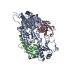
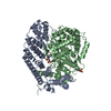
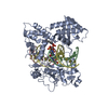
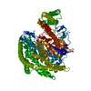
| Title | Structural basis for substrate recognition by a novel Legionella phosphoinositide phosphatase | ||||||
 Components Components | SidF, inhibitor of growth family, member 3 | ||||||
 Keywords Keywords | HYDROLASE / mixed alpha-beta / phosphoinositide phosphatase / phosphoinositides / membrane | ||||||
| Function / homology | : / Phosphoinositide phosphatase / phosphatidylinositol phosphate phosphatase activity / Chem-3PT / SidF, inhibitor of growth family, member 3 Function and homology information Function and homology information | ||||||
| Biological species |  Legionella pneumophila subsp. pneumophila (bacteria) Legionella pneumophila subsp. pneumophila (bacteria) | ||||||
| Method |  X-RAY DIFFRACTION / SYNCHROTRON / FOURIER SYNTHESIS / Resolution: 2.822 Å X-RAY DIFFRACTION / SYNCHROTRON / FOURIER SYNTHESIS / Resolution: 2.822 Å | ||||||
 Authors Authors | Hsu, F.S. / Zhu, W. / Brennan, L. / Tao, L. / Luo, Z.Q. / Mao, Y. | ||||||
 Citation Citation | Journal: Proc.Natl.Acad.Sci.USA / Year: 2012 Title: Structural basis for substrate recognition by a unique Legionella phosphoinositide phosphatase. Authors: Hsu, F. / Zhu, W. / Brennan, L. / Tao, L. / Luo, Z.Q. / Mao, Y. | ||||||
| History |
|
- Structure visualization
| Structure viewer | Molecule: MolmilJmol/JSmol |
|---|
- Downloads & links
-Download
| PDBx/mmCIF format | 4fyg.cif.gz | 308.9 KB | Display | PDBx/mmCIF format |
|---|---|---|---|---|
| PDB format | pdb4fyg.ent.gz | 253 KB | Display | PDB format |
| PDBx/mmJSON format | 4fyg.json.gz | Tree view | PDBx/mmJSON format | |
| Others |  Other downloads Other downloads |
-Validation report
| Arichive directory | https://data.pdbj.org/pub/pdb/validation_reports/fy/4fygftp://data.pdbj.org/pub/pdb/validation_reports/fy/4fyg | HTTPS FTP |
|---|
-Related structure data
| Related structure data |  4fyeC  4fyfSC C: citing same article ( S: Starting model for refinement |
|---|---|
| Similar structure data |


-Links
 PDBj
PDBj- Assembly
| Deposited unit | 
| ||||||||
|---|---|---|---|---|---|---|---|---|---|
| 1 |
| ||||||||
| Unit cell |
|
-Components
| #1: Protein | Mass: 86191.359 Da / Num. of mol.: 1 Fragment: N-terminal phosphatase domain of SidF (UNP Residues 1-760) Mutation: C645S Source method: isolated from a genetically manipulated source Source: (gene. exp.) Legionella pneumophila subsp. pneumophila (bacteria)Strain: Philadelphia 1 / ATCC 33152 / DSM 7513 / Gene: lpg2584, sidF / Plasmid: Pet28a / Production host: References: UniProt: Q5ZSD5, phosphatidylinositol-3,4,5-trisphosphate 3-phosphatase |
|---|---|
| #2: Chemical | ChemComp-3PT / (  Mass: 634.354 Da / Num. of mol.: 1 / Source method: obtained synthetically / Formula: C17H33O19P3 Mass: 634.354 Da / Num. of mol.: 1 / Source method: obtained synthetically / Formula: C17H33O19P3 |
| #3: Water | ChemComp-HOH /  Mass: 18.015 Da / Num. of mol.: 6 / Source method: isolated from a natural source / Formula: H2O Mass: 18.015 Da / Num. of mol.: 6 / Source method: isolated from a natural source / Formula: H2O |
-Experimental details
-Experiment
| Experiment | Method: X-RAY DIFFRACTION / Number of used crystals: 1 |
|---|
- Sample preparation
| Crystal | Density Matthews: 2.95 Å3/Da / Density % sol: 58.31 % |
|---|---|
| Crystal grow | Temperature: 298 K / Method: vapor diffusion, hanging drop / pH: 8 Details: 0.1 M Tris-HCl pH8.0, 10% PEG3350, and 10% ethylene glycol, VAPOR DIFFUSION, HANGING DROP, temperature 298K |
-Data collection
| Diffraction | Mean temperature: 100 K |
|---|---|
| Diffraction source | Source: SYNCHROTRON / Site: NSLS  / Beamline: X4C / Wavelength: 0.9789 Å / Beamline: X4C / Wavelength: 0.9789 Å |
| Detector | Type: MAR CCD 165 mm / Detector: CCD / Date: Feb 4, 2012 / Details: mirrors |
| Radiation | Monochromator: Single crystal bender / Protocol: SINGLE WAVELENGTH / Monochromatic (M) / Laue (L): M / Scattering type: x-ray |
| Radiation wavelength | Wavelength: 0.9789 Å / Relative weight: 1 |
| Reflection | Resolution: 2.82→50 Å / Num. all: 301119 / Num. obs: 24387 / % possible obs: 96.8 % / Observed criterion σ(F): 1 / Observed criterion σ(I): 1 / Redundancy: 12.3 % / Rmerge(I) obs: 0.102 / Net I/σ(I): 28.6 |
| Reflection shell | Resolution: 2.82→2.9 Å / % possible all: 78.9 |
- Processing
| Software |
| |||||||||||||||||||||||||||||||||||||||||||||
|---|---|---|---|---|---|---|---|---|---|---|---|---|---|---|---|---|---|---|---|---|---|---|---|---|---|---|---|---|---|---|---|---|---|---|---|---|---|---|---|---|---|---|---|---|---|---|
| Refinement | Method to determine structure: FOURIER SYNTHESIS Starting model: PDB entry 4FYF Resolution: 2.822→36.58 Å / Cor.coef. Fo:Fc: 0.958 / Cor.coef. Fo:Fc free: 0.91 / SU B: 30.281 / SU ML: 0.272 / Cross valid method: THROUGHOUT / ESU R Free: 0.375 / Stereochemistry target values: MAXIMUM LIKELIHOOD / Details: HYDROGENS HAVE BEEN USED IF PRESENT IN THE INPUT
| |||||||||||||||||||||||||||||||||||||||||||||
| Solvent computation | Ion probe radii: 0.8 Å / Shrinkage radii: 0.8 Å / VDW probe radii: 1.2 Å / Solvent model: MASK | |||||||||||||||||||||||||||||||||||||||||||||
| Displacement parameters | Biso mean: 70.396 Å2
| |||||||||||||||||||||||||||||||||||||||||||||
| Refinement step | Cycle: LAST / Resolution: 2.822→36.58 Å
| |||||||||||||||||||||||||||||||||||||||||||||
| Refine LS restraints |
| |||||||||||||||||||||||||||||||||||||||||||||
| LS refinement shell | Resolution: 2.822→2.895 Å / Total num. of bins used: 20
| |||||||||||||||||||||||||||||||||||||||||||||
| Refinement TLS params. | Method: refined / Origin x: -2.5584 Å / Origin y: 45.7343 Å / Origin z: 21.9739 Å
|
