| Entry | Database: PDB / ID: 4jyk
|
|---|
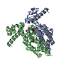
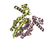
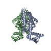
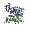
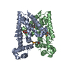
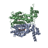
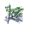
| Title | Structure of E. coli Transcriptional Regulator RutR with bound uracil |
|---|
 Components Components | HTH-type transcriptional regulator RutR |
|---|
 Keywords Keywords | TRANSCRIPTION REGULATOR / Structural Genomics / PSI-Biology / New York Structural Genomics Research Consortium / NYSGRC / Helix-turn-Helix / Transcriptional Regulator |
|---|
| Function / homology |  Function and homology information Function and homology information
transcription cis-regulatory region binding / DNA-binding transcription factor activity / negative regulation of DNA-templated transcription / regulation of DNA-templated transcription / positive regulation of DNA-templated transcriptionSimilarity search - Function Transcription regulator YcdC, C-terminal / Transcription regulator, pyrimidine utilisation, RutR / YcdC-like protein, C-terminal region / : / Tetracycline Repressor, domain 2 / Tetracyclin repressor-like, C-terminal domain superfamily / Tetracycline Repressor; domain 2 / Bacterial regulatory proteins, tetR family / DNA-binding HTH domain, TetR-type / TetR-type HTH domain profile. ...Transcription regulator YcdC, C-terminal / Transcription regulator, pyrimidine utilisation, RutR / YcdC-like protein, C-terminal region / : / Tetracycline Repressor, domain 2 / Tetracyclin repressor-like, C-terminal domain superfamily / Tetracycline Repressor; domain 2 / Bacterial regulatory proteins, tetR family / DNA-binding HTH domain, TetR-type / TetR-type HTH domain profile. / Homeodomain-like / Homeobox-like domain superfamily / Arc Repressor Mutant, subunit A / Orthogonal Bundle / Mainly AlphaSimilarity search - Domain/homology |
|---|
| Biological species |   Escherichia coli (E. coli) Escherichia coli (E. coli) |
|---|
| Method |  X-RAY DIFFRACTION / SYNCHROTRON / molecular replacement / Resolution: 1.7 Å X-RAY DIFFRACTION / SYNCHROTRON / molecular replacement / Resolution: 1.7 Å |
|---|
 Authors Authors | Cooper, D.R. / Knapik, A.A. / Petkowski, J.J. / Porebski, P.J. / Osinski, T. / Almo, S.C. / Minor, W. / New York Structural Genomics Research Consortium (NYSGRC) |
|---|
 Citation Citation | Journal: To be Published
Title: Structure of E. coli Transcriptional Regulator RutR with bound uracil
Authors: Cooper, D.R. / Knapik, A.A. / Petkowski, J.J. / Porebski, P.J. / Osinski, T. / Almo, S.C. / Minor, W. / New York Structural Genomics Research Consortium (NYSGRC) |
|---|
| History | | Deposition | Mar 29, 2013 | Deposition site: RCSB / Processing site: RCSB |
|---|
| Revision 1.0 | Aug 14, 2013 | Provider: repository / Type: Initial release |
|---|
| Revision 1.1 | Nov 15, 2017 | Group: Refinement description / Category: software |
|---|
| Revision 1.2 | Apr 13, 2022 | Group: Database references / Derived calculations / Structure summary
Category: audit_author / citation_author ...audit_author / citation_author / database_2 / struct_site
Item: _audit_author.identifier_ORCID / _citation_author.identifier_ORCID ..._audit_author.identifier_ORCID / _citation_author.identifier_ORCID / _database_2.pdbx_DOI / _database_2.pdbx_database_accession / _struct_site.pdbx_auth_asym_id / _struct_site.pdbx_auth_comp_id / _struct_site.pdbx_auth_seq_id |
|---|
| Revision 1.3 | Feb 28, 2024 | Group: Data collection / Refinement description
Category: chem_comp_atom / chem_comp_bond / struct_ncs_dom_lim
Item: _struct_ncs_dom_lim.beg_auth_comp_id / _struct_ncs_dom_lim.beg_label_asym_id ..._struct_ncs_dom_lim.beg_auth_comp_id / _struct_ncs_dom_lim.beg_label_asym_id / _struct_ncs_dom_lim.beg_label_comp_id / _struct_ncs_dom_lim.beg_label_seq_id / _struct_ncs_dom_lim.end_auth_comp_id / _struct_ncs_dom_lim.end_label_asym_id / _struct_ncs_dom_lim.end_label_comp_id / _struct_ncs_dom_lim.end_label_seq_id |
|---|
|
|---|
 Movie
Movie Controller

 Yorodumi
 Open data
 Basic information
 Structure visualization
 Downloads & links
 Other downloads
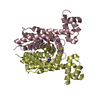


 PDBj
PDBj Assembly
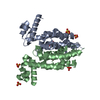

 Mass: 96.063 Da / Num. of mol.: 7 / Source method: obtained synthetically / Formula: SO4
 Mass: 112.087 Da / Num. of mol.: 2 / Source method: obtained synthetically / Formula: C4H4N2O2
 Mass: 18.015 Da / Num. of mol.: 279 / Source method: isolated from a natural source / Formula: H2O
 Sample preparation
 / Beamline: 21-ID-G / Wavelength: 0.97856 Å
 Processing