Mass: 18.015 Da / Num. of mol.: 35 / Source method: isolated from a natural source / Formula: H2O
-
Details
Has protein modification
Y
-
Experimental details
-
Experiment
Experiment
Method: X-RAY DIFFRACTION / Number of used crystals: 1
-
Sample preparation
Crystal
Density Matthews: 4.14 Å3/Da / Density % sol: 70.31 %
Crystal grow
Temperature: 277 K / Method: vapor diffusion, hanging drop / pH: 3.5 Details: 28% PEG400, 0.1 M citric acid pH 3.5, 0.1M NaCl and 0.1M Li2SO4. The crystal was subsequently soaked with 50mM NaNO2 in the above solution prior to freezing, VAPOR DIFFUSION, HANGING DROP, temperature 277K
-
Data collection
Diffraction
Mean temperature: 100 K
Diffraction source
Source: SYNCHROTRON / Site: ALS / Beamline: 8.2.2 / Wavelength: 0.99999 Å
Resolution: 2.8→50 Å / Cor.coef. Fo:Fc: 0.889 / Cor.coef. Fo:Fc free: 0.859 / SU B: 12.544 / SU ML: 0.247 / Isotropic thermal model: Isotropic / Cross valid method: THROUGHOUT / σ(F): 0 / ESU R: 0.54 / ESU R Free: 0.334 / Stereochemistry target values: MAXIMUM LIKELIHOOD Details: A strong density on the surface of NarK is likely from the sugar head group of the decylmaltoside detergent used during protein purification and is modelled in the structure as maltose.
Rfactor
Num. reflection
% reflection
Selection details
Rfree
0.2686
3642
5 %
RANDOM
Rwork
0.22337
-
-
-
obs
0.22567
68494
94.53 %
-
all
-
72136
-
-
Solvent computation
Ion probe radii: 0.8 Å / Shrinkage radii: 0.8 Å / VDW probe radii: 1.2 Å / Solvent model: MASK
Displacement parameters
Biso mean: 57.172 Å2
Baniso -1
Baniso -2
Baniso -3
1-
-0.68 Å2
-0.23 Å2
-1.11 Å2
2-
-
-0.1 Å2
1.65 Å2
3-
-
-
0.91 Å2
Refine analyze
Luzzati coordinate error obs: 0.3741 Å
Refinement step
Cycle: LAST / Resolution: 2.8→50 Å
Protein
Nucleic acid
Ligand
Solvent
Total
Num. atoms
12522
0
32
35
12589
Refine LS restraints
Refine-ID
Type
Dev ideal
Dev ideal target
Number
X-RAY DIFFRACTION
r_bond_refined_d
0.008
0.02
12889
X-RAY DIFFRACTION
r_angle_refined_deg
1.29
1.946
17560
X-RAY DIFFRACTION
r_dihedral_angle_1_deg
6.018
5
1646
X-RAY DIFFRACTION
r_dihedral_angle_2_deg
37.015
23.137
459
X-RAY DIFFRACTION
r_dihedral_angle_3_deg
20.783
15
1921
X-RAY DIFFRACTION
r_dihedral_angle_4_deg
17.516
15
46
X-RAY DIFFRACTION
r_chiral_restr
0.085
0.2
2011
X-RAY DIFFRACTION
r_gen_planes_refined
0.005
0.021
9620
LS refinement shell
Resolution: 2.8→2.873 Å / Total num. of bins used: 20
Rfactor
Num. reflection
% reflection
Rfree
0.37
223
-
Rwork
0.312
4088
-
obs
-
4088
77.26 %
+
About Yorodumi
-
News
-
Feb 9, 2022. New format data for meta-information of EMDB entries
New format data for meta-information of EMDB entries
Version 3 of the EMDB header file is now the official format.
The previous official version 1.9 will be removed from the archive.
In the structure databanks used in Yorodumi, some data are registered as the other names, "COVID-19 virus" and "2019-nCoV". Here are the details of the virus and the list of structure data.
Jan 31, 2019. EMDB accession codes are about to change! (news from PDBe EMDB page)
EMDB accession codes are about to change! (news from PDBe EMDB page)
The allocation of 4 digits for EMDB accession codes will soon come to an end. Whilst these codes will remain in use, new EMDB accession codes will include an additional digit and will expand incrementally as the available range of codes is exhausted. The current 4-digit format prefixed with “EMD-” (i.e. EMD-XXXX) will advance to a 5-digit format (i.e. EMD-XXXXX), and so on. It is currently estimated that the 4-digit codes will be depleted around Spring 2019, at which point the 5-digit format will come into force.
The EM Navigator/Yorodumi systems omit the EMD- prefix.
Related info.:Q: What is EMD? / ID/Accession-code notation in Yorodumi/EM Navigator
Yorodumi is a browser for structure data from EMDB, PDB, SASBDB, etc.
This page is also the successor to EM Navigator detail page, and also detail information page/front-end page for Omokage search.
The word "yorodu" (or yorozu) is an old Japanese word meaning "ten thousand". "mi" (miru) is to see.
Related info.:EMDB / PDB / SASBDB / Comparison of 3 databanks / Yorodumi Search / Aug 31, 2016. New EM Navigator & Yorodumi / Yorodumi Papers / Jmol/JSmol / Function and homology information / Changes in new EM Navigator and Yorodumi
 Movie
Movie Controller

 Yorodumi
 Open data
 Basic information
 Components
 Keywords
 Function and homology information

 X-RAY DIFFRACTION /
  Authors
 Citation
 Structure visualization
 Downloads & links
 Other downloads
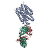


 PDBj
PDBj


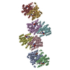
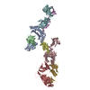
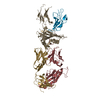
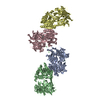
 Assembly
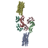

 Type: D-saccharide / Mass: 194.182 Da / Num. of mol.: 2
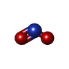

 Mass: 46.005 Da / Num. of mol.: 2 / Source method: obtained synthetically / Formula: NO2
 Sample preparation
 / Beamline: 8.2.2 / Wavelength: 0.99999 Å
 Processing