 Movie
Movie Controller

[English] 日本語
 Yorodumi
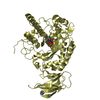
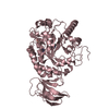
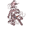
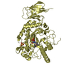
- PDB-4jfu: Crystal structure of a bacterial fucosidase with iminosugar inhibitor -
+ Open data

- Basic information
| Entry | Database: PDB / ID: 4jfu | ||||||
|---|---|---|---|---|---|---|---|
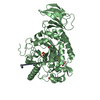
| Title | Crystal structure of a bacterial fucosidase with iminosugar inhibitor | ||||||
 Components Components | alpha-L-fucosidase | ||||||
 Keywords Keywords | HYDROLASE/HYDROLASE INHIBITOR / alpha-L-fucosidase / HYDROLASE-HYDROLASE INHIBITOR complex | ||||||
| Function / homology |  Function and homology information Function and homology informationalpha-L-fucosidase / alpha-L-fucosidase activity / fucose metabolic process / glycoside catabolic process / lysosome Similarity search - Function | ||||||
| Biological species |  Bacteroides thetaiotaomicron (bacteria) Bacteroides thetaiotaomicron (bacteria) | ||||||
| Method |  X-RAY DIFFRACTION / SYNCHROTRON / MOLECULAR REPLACEMENT / Resolution: 1.66 Å X-RAY DIFFRACTION / SYNCHROTRON / MOLECULAR REPLACEMENT / Resolution: 1.66 Å | ||||||
 Authors Authors | Wright, D.W. / Davies, G.J. | ||||||
 Citation Citation | Journal: Chemistry / Year: 2013 Title: alpha-L-fucosidase inhibition by pyrrolidine-ferrocene hybrids: rationalization of ligand-binding properties by structural studies. Authors: Hottin, A. / Wright, D.W. / Steenackers, A. / Delannoy, P. / Dubar, F. / Biot, C. / Davies, G.J. / Behr, J.B. | ||||||
| History |
|
- Structure visualization
| Structure viewer | Molecule: MolmilJmol/JSmol |
|---|
- Downloads & links
-Download
| PDBx/mmCIF format | 4jfu.cif.gz | 218.4 KB | Display | PDBx/mmCIF format |
|---|---|---|---|---|
| PDB format | pdb4jfu.ent.gz | 170.2 KB | Display | PDB format |
| PDBx/mmJSON format | 4jfu.json.gz | Tree view | PDBx/mmJSON format | |
| Others |  Other downloads Other downloads |
-Validation report
| Arichive directory | https://data.pdbj.org/pub/pdb/validation_reports/jf/4jfuftp://data.pdbj.org/pub/pdb/validation_reports/jf/4jfu | HTTPS FTP |
|---|
-Related structure data
| Related structure data |  4jfsC  4jftC  4jfvC  4jfwC  4j27S S: Starting model for refinement C: citing same article ( |
|---|---|
| Similar structure data |


-Links
 PDBj
PDBj


- Assembly
| Deposited unit | 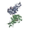
| ||||||||
|---|---|---|---|---|---|---|---|---|---|
| 1 | 
| ||||||||
| 2 | 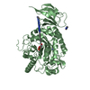
| ||||||||
| Unit cell |
|
-Components
-Protein , 1 types, 2 molecules AB
| #1: Protein | Mass: 52080.930 Da / Num. of mol.: 2 / Fragment: RESIDUES 35-484 Source method: isolated from a genetically manipulated source Source: (gene. exp.) Bacteroides thetaiotaomicron (bacteria)Strain: VPI-5482 / Plasmid: pET-YSBLIC / Production host: |
|---|
-Non-polymers , 5 types, 1113 molecules 


| #2: Chemical |  Mass: 207.269 Da / Num. of mol.: 2 / Source method: obtained synthetically / Formula: C12H17NO2 Mass: 207.269 Da / Num. of mol.: 2 / Source method: obtained synthetically / Formula: C12H17NO2#3: Chemical |  Mass: 96.063 Da / Num. of mol.: 3 / Source method: obtained synthetically / Formula: SO4 Mass: 96.063 Da / Num. of mol.: 3 / Source method: obtained synthetically / Formula: SO4#4: Chemical | ChemComp-IMD /  Mass: 69.085 Da / Num. of mol.: 5 / Source method: obtained synthetically / Formula: C3H5N2 Mass: 69.085 Da / Num. of mol.: 5 / Source method: obtained synthetically / Formula: C3H5N2#5: Chemical | ChemComp-GOL /  Mass: 92.094 Da / Num. of mol.: 5 / Source method: obtained synthetically / Formula: C3H8O3 Mass: 92.094 Da / Num. of mol.: 5 / Source method: obtained synthetically / Formula: C3H8O3#6: Water | ChemComp-HOH / | Mass: 18.015 Da / Num. of mol.: 1098 / Source method: isolated from a natural source / Formula: H2O |
|---|
-Experimental details
-Experiment
| Experiment | Method: X-RAY DIFFRACTION / Number of used crystals: 1 |
|---|
- Sample preparation
| Crystal | Density Matthews: 3.02 Å3/Da / Density % sol: 59.26 % |
|---|---|
| Crystal grow | Temperature: 291.5 K / Method: vapor diffusion, hanging drop / pH: 7 Details: 0.16 M ammonium sulfate, 20% PEG 6K, 0.1M imidazole pH 7, VAPOR DIFFUSION, HANGING DROP, temperature 291.5K |
-Data collection
| Diffraction | Mean temperature: 100 K | ||||||||||||||||||||||||||||||||||||||||||||||||||||||||||||||||||||||||||||||||||||||||
|---|---|---|---|---|---|---|---|---|---|---|---|---|---|---|---|---|---|---|---|---|---|---|---|---|---|---|---|---|---|---|---|---|---|---|---|---|---|---|---|---|---|---|---|---|---|---|---|---|---|---|---|---|---|---|---|---|---|---|---|---|---|---|---|---|---|---|---|---|---|---|---|---|---|---|---|---|---|---|---|---|---|---|---|---|---|---|---|---|---|
| Diffraction source | Source: SYNCHROTRON / Site: Diamond  / Beamline: I03 / Wavelength: 0.9763 Å / Beamline: I03 / Wavelength: 0.9763 Å | ||||||||||||||||||||||||||||||||||||||||||||||||||||||||||||||||||||||||||||||||||||||||
| Detector | Type: PSI PILATUS 6M / Detector: PIXEL / Date: Dec 12, 2011 | ||||||||||||||||||||||||||||||||||||||||||||||||||||||||||||||||||||||||||||||||||||||||
| Radiation | Monochromator: Si 111 CHANNEL / Protocol: SINGLE WAVELENGTH / Monochromatic (M) / Laue (L): M / Scattering type: x-ray | ||||||||||||||||||||||||||||||||||||||||||||||||||||||||||||||||||||||||||||||||||||||||
| Radiation wavelength | Wavelength: 0.9763 Å / Relative weight: 1 | ||||||||||||||||||||||||||||||||||||||||||||||||||||||||||||||||||||||||||||||||||||||||
| Reflection | Resolution: 1.659→55.385 Å / Num. all: 145075 / Num. obs: 145075 / % possible obs: 99.3 % / Observed criterion σ(F): 0 / Observed criterion σ(I): 0 / Redundancy: 2.9 % / Rsym value: 0.047 / Net I/σ(I): 10.7 | ||||||||||||||||||||||||||||||||||||||||||||||||||||||||||||||||||||||||||||||||||||||||
| Reflection shell | Diffraction-ID: 1
|
- Processing
| Software |
| ||||||||||||||||||||||||||||||||||||||||||||||||||||||||||||
|---|---|---|---|---|---|---|---|---|---|---|---|---|---|---|---|---|---|---|---|---|---|---|---|---|---|---|---|---|---|---|---|---|---|---|---|---|---|---|---|---|---|---|---|---|---|---|---|---|---|---|---|---|---|---|---|---|---|---|---|---|---|
| Refinement | Method to determine structure: MOLECULAR REPLACEMENT Starting model: 4J27 Resolution: 1.66→55.385 Å / Cor.coef. Fo:Fc: 0.976 / Cor.coef. Fo:Fc free: 0.968 / WRfactor Rfree: 0.1929 / WRfactor Rwork: 0.1629 / Occupancy max: 1 / Occupancy min: 0 / FOM work R set: 0.8775 / SU B: 1.638 / SU ML: 0.054 / SU R Cruickshank DPI: 0.073 / SU Rfree: 0.0741 / Cross valid method: THROUGHOUT / σ(F): 0 / ESU R: 0.073 / ESU R Free: 0.074 / Stereochemistry target values: MAXIMUM LIKELIHOOD Details: HYDROGENS HAVE BEEN USED IF PRESENT IN THE INPUT U VALUES: REFINED INDIVIDUALLY. THE MOLECULAR REPLACEMENT WAS NOT USED, COORDINATES FROM 4J27 WERE CONVERTED INTO THE (ALMOST) ISOMORPHOUS ...Details: HYDROGENS HAVE BEEN USED IF PRESENT IN THE INPUT U VALUES: REFINED INDIVIDUALLY. THE MOLECULAR REPLACEMENT WAS NOT USED, COORDINATES FROM 4J27 WERE CONVERTED INTO THE (ALMOST) ISOMORPHOUS CELL OF 4JFU TO BUILD A STARTING MODEL DIRECTLY. THE RFREE FLAG FROM 4J27 WAS USED FOR THIS DATASET.
| ||||||||||||||||||||||||||||||||||||||||||||||||||||||||||||
| Solvent computation | Ion probe radii: 0.8 Å / Shrinkage radii: 0.8 Å / VDW probe radii: 1.2 Å / Solvent model: MASK | ||||||||||||||||||||||||||||||||||||||||||||||||||||||||||||
| Displacement parameters | Biso max: 75.72 Å2 / Biso mean: 26.2193 Å2 / Biso min: 14.42 Å2
| ||||||||||||||||||||||||||||||||||||||||||||||||||||||||||||
| Refinement step | Cycle: LAST / Resolution: 1.66→55.385 Å
| ||||||||||||||||||||||||||||||||||||||||||||||||||||||||||||
| Refine LS restraints |
| ||||||||||||||||||||||||||||||||||||||||||||||||||||||||||||
| LS refinement shell | Resolution: 1.66→1.702 Å / Total num. of bins used: 20
|
