 Movie
Movie Controller

[English] 日本語
 Yorodumi
- PDB-4jfd: Preservation of peptide specificity during TCR-MHC contact domina... -
+ Open data

- Basic information
| Entry | Database: PDB / ID: 4jfd | ||||||
|---|---|---|---|---|---|---|---|
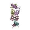
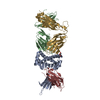
| Title | Preservation of peptide specificity during TCR-MHC contact dominated affinity enhancement of a melanoma-specific TCR | ||||||
 Components Components |
| ||||||
 Keywords Keywords | IMMUNE SYSTEM / HLA / TCR / Melanoma / High Affinity / Immunoglobulin | ||||||
| Function / homology |  Function and homology information Function and homology informationalpha-beta T cell receptor complex / positive regulation of memory T cell activation / T cell mediated cytotoxicity directed against tumor cell target / positive regulation of CD8-positive, alpha-beta T cell activation / CD8-positive, alpha-beta T cell activation / positive regulation of CD8-positive, alpha-beta T cell proliferation / antigen processing and presentation of endogenous peptide antigen via MHC class I via ER pathway, TAP-dependent / TAP complex binding / antigen processing and presentation of exogenous peptide antigen via MHC class I / Golgi medial cisterna ...alpha-beta T cell receptor complex / positive regulation of memory T cell activation / T cell mediated cytotoxicity directed against tumor cell target / positive regulation of CD8-positive, alpha-beta T cell activation / CD8-positive, alpha-beta T cell activation / positive regulation of CD8-positive, alpha-beta T cell proliferation / antigen processing and presentation of endogenous peptide antigen via MHC class I via ER pathway, TAP-dependent / TAP complex binding / antigen processing and presentation of exogenous peptide antigen via MHC class I / Golgi medial cisterna / Translocation of ZAP-70 to Immunological synapse / Phosphorylation of CD3 and TCR zeta chains / CD8 receptor binding / protection from natural killer cell mediated cytotoxicity / alpha-beta T cell activation / Generation of second messenger molecules / beta-2-microglobulin binding / endoplasmic reticulum exit site / Co-inhibition by PD-1 / TAP binding / detection of bacterium / antigen processing and presentation of endogenous peptide antigen via MHC class Ib / antigen processing and presentation of endogenous peptide antigen via MHC class I via ER pathway, TAP-independent / T cell receptor binding / early endosome lumen / Nef mediated downregulation of MHC class I complex cell surface expression / DAP12 interactions / Endosomal/Vacuolar pathway / T cell mediated cytotoxicity / response to bacterium / Antigen Presentation: Folding, assembly and peptide loading of class I MHC / lumenal side of endoplasmic reticulum membrane / regulation of iron ion transport / cellular response to iron(III) ion / negative regulation of iron ion transport / negative regulation of forebrain neuron differentiation / antigen processing and presentation of exogenous protein antigen via MHC class Ib, TAP-dependent / peptide antigen assembly with MHC class I protein complex / ER to Golgi transport vesicle membrane / regulation of erythrocyte differentiation / response to molecule of bacterial origin / HFE-transferrin receptor complex / MHC class I peptide loading complex / transferrin transport / cellular response to iron ion / negative regulation of receptor-mediated endocytosis / positive regulation of T cell cytokine production / antigen processing and presentation of endogenous peptide antigen via MHC class I / MHC class I protein complex / peptide antigen assembly with MHC class II protein complex / negative regulation of neurogenesis / MHC class II protein complex / cellular response to nicotine / positive regulation of receptor-mediated endocytosis / multicellular organismal-level iron ion homeostasis / positive regulation of T cell mediated cytotoxicity / specific granule lumen / antigen processing and presentation of exogenous peptide antigen via MHC class II / positive regulation of immune response / peptide antigen binding / positive regulation of type II interferon production / phagocytic vesicle membrane / recycling endosome membrane / positive regulation of T cell activation / negative regulation of epithelial cell proliferation / Interferon gamma signaling / Immunoregulatory interactions between a Lymphoid and a non-Lymphoid cell / Interferon alpha/beta signaling / sensory perception of smell / Modulation by Mtb of host immune system / positive regulation of cellular senescence / tertiary granule lumen / MHC class II protein complex binding / T cell differentiation in thymus / DAP12 signaling / late endosome membrane / Downstream TCR signaling / negative regulation of neuron projection development / T cell receptor signaling pathway / antibacterial humoral response / E3 ubiquitin ligases ubiquitinate target proteins / protein refolding / ER-Phagosome pathway / early endosome membrane / amyloid fibril formation / protein homotetramerization / intracellular iron ion homeostasis / adaptive immune response / learning or memory / defense response to Gram-positive bacterium / immune response / endoplasmic reticulum lumen / Amyloid fiber formation / signaling receptor binding / Golgi membrane / external side of plasma membrane / innate immune response / lysosomal membrane / focal adhesion / Neutrophil degranulation Similarity search - Function | ||||||
| Biological species |  Homo sapiens (human) Homo sapiens (human) | ||||||
| Method |  X-RAY DIFFRACTION / SYNCHROTRON / MOLECULAR REPLACEMENT / molecular replacement / Resolution: 2.46 Å X-RAY DIFFRACTION / SYNCHROTRON / MOLECULAR REPLACEMENT / molecular replacement / Resolution: 2.46 Å | ||||||
 Authors Authors | Rizkallah, P.J. / Cole, D.K. / Madura, F. / Sewell, A.K. | ||||||
 Citation Citation | Journal: J.Biol.Chem. / Year: 2013 Title: T-cell receptor specificity maintained by altered thermodynamics. Authors: Madura, F. / Rizkallah, P.J. / Miles, K.M. / Holland, C.J. / Bulek, A.M. / Fuller, A. / Schauenburg, A.J. / Miles, J.J. / Liddy, N. / Sami, M. / Li, Y. / Hossain, M. / Baker, B.M. / ...Authors: Madura, F. / Rizkallah, P.J. / Miles, K.M. / Holland, C.J. / Bulek, A.M. / Fuller, A. / Schauenburg, A.J. / Miles, J.J. / Liddy, N. / Sami, M. / Li, Y. / Hossain, M. / Baker, B.M. / Jakobsen, B.K. / Sewell, A.K. / Cole, D.K. | ||||||
| History |
|
- Structure visualization
| Structure viewer | Molecule: MolmilJmol/JSmol |
|---|
- Downloads & links
-Download
| PDBx/mmCIF format | 4jfd.cif.gz | 355.5 KB | Display | PDBx/mmCIF format |
|---|---|---|---|---|
| PDB format | pdb4jfd.ent.gz | 290.5 KB | Display | PDB format |
| PDBx/mmJSON format | 4jfd.json.gz | Tree view | PDBx/mmJSON format | |
| Others |  Other downloads Other downloads |
-Validation report
| Arichive directory | https://data.pdbj.org/pub/pdb/validation_reports/jf/4jfdftp://data.pdbj.org/pub/pdb/validation_reports/jf/4jfd | HTTPS FTP |
|---|
-Related structure data
| Related structure data |  4jfeC  4jffC  4jfhC 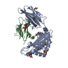 4jfoC 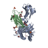 4jfpC 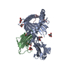 4jfqC C: citing same article ( |
|---|---|
| Similar structure data |


-Links
 PDBj
PDBj


- Assembly
| Deposited unit | 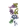
| ||||||||
|---|---|---|---|---|---|---|---|---|---|
| 1 |
| ||||||||
| Unit cell |
|
-Components
-Protein , 2 types, 2 molecules AB
| #1: Protein | Mass: 31951.316 Da / Num. of mol.: 1 Source method: isolated from a genetically manipulated source Source: (gene. exp.) Homo sapiens (human) / Gene: HLA-A, HLAA / Plasmid: pGMT7 / Production host:  |
|---|---|
| #2: Protein | Mass: 11879.356 Da / Num. of mol.: 1 Source method: isolated from a genetically manipulated source Source: (gene. exp.) Homo sapiens (human) / Gene: B2M, CDABP0092, HDCMA22P / Plasmid: pGMT7 / Production host: |
-Protein/peptide , 1 types, 1 molecules C
| #3: Protein/peptide | Mass: 999.202 Da / Num. of mol.: 1 / Source method: obtained synthetically / Details: Heteroclitic G4A version of Melanoma epitope |
|---|
-High Affinity TCR ... , 2 types, 2 molecules DE
| #4: Protein | Mass: 21739.879 Da / Num. of mol.: 1 Source method: isolated from a genetically manipulated source Source: (gene. exp.) Homo sapiens (human) / Plasmid: pGMT7 / Production host: |
|---|---|
| #5: Protein | Mass: 27522.686 Da / Num. of mol.: 1 Source method: isolated from a genetically manipulated source Source: (gene. exp.) Homo sapiens (human) / Plasmid: pGMT7 / Production host: |
-Non-polymers , 3 types, 137 molecules 


| #6: Chemical | ChemComp-TAM /  Mass: 163.215 Da / Num. of mol.: 1 / Source method: obtained synthetically / Formula: C7H17NO3 / Comment: pH buffer*YM Mass: 163.215 Da / Num. of mol.: 1 / Source method: obtained synthetically / Formula: C7H17NO3 / Comment: pH buffer*YM | ||
|---|---|---|---|
| #7: Chemical | ChemComp-SO4 /  Mass: 96.063 Da / Num. of mol.: 11 / Source method: obtained synthetically / Formula: SO4 Mass: 96.063 Da / Num. of mol.: 11 / Source method: obtained synthetically / Formula: SO4#8: Water | ChemComp-HOH / | Mass: 18.015 Da / Num. of mol.: 125 / Source method: isolated from a natural source / Formula: H2O |
-Details
| Has protein modification | Y |
|---|
-Experimental details
-Experiment
| Experiment | Method: X-RAY DIFFRACTION / Number of used crystals: 1 |
|---|
- Sample preparation
| Crystal | Density Matthews: 3.25 Å3/Da / Density % sol: 62.19 % |
|---|---|
| Crystal grow | Temperature: 291 K / Method: vapor diffusion, sitting drop / pH: 7.5 Details: 0.1 M TRIS, 15% PEG 4000, 17.4% glycerol, pH 7.5, vapor diffusion, sitting drop, temperature 291K |
-Data collection
| Diffraction | Mean temperature: 100 K | ||||||||||||||||||||||||||||||||||||||||||||||||||||||||||||||||||||||||||||||||||||||||||||||||||||||||||||||||||||||||||||||||||||||||||||||||||||||||||||||||||||||||
|---|---|---|---|---|---|---|---|---|---|---|---|---|---|---|---|---|---|---|---|---|---|---|---|---|---|---|---|---|---|---|---|---|---|---|---|---|---|---|---|---|---|---|---|---|---|---|---|---|---|---|---|---|---|---|---|---|---|---|---|---|---|---|---|---|---|---|---|---|---|---|---|---|---|---|---|---|---|---|---|---|---|---|---|---|---|---|---|---|---|---|---|---|---|---|---|---|---|---|---|---|---|---|---|---|---|---|---|---|---|---|---|---|---|---|---|---|---|---|---|---|---|---|---|---|---|---|---|---|---|---|---|---|---|---|---|---|---|---|---|---|---|---|---|---|---|---|---|---|---|---|---|---|---|---|---|---|---|---|---|---|---|---|---|---|---|---|---|---|---|
| Diffraction source | Source: SYNCHROTRON / Site: Diamond  / Beamline: I03 / Wavelength: 0.9763 Å / Beamline: I03 / Wavelength: 0.9763 Å | ||||||||||||||||||||||||||||||||||||||||||||||||||||||||||||||||||||||||||||||||||||||||||||||||||||||||||||||||||||||||||||||||||||||||||||||||||||||||||||||||||||||||
| Detector | Detector: CCD / Date: Oct 8, 2010 / Details: mirrors | ||||||||||||||||||||||||||||||||||||||||||||||||||||||||||||||||||||||||||||||||||||||||||||||||||||||||||||||||||||||||||||||||||||||||||||||||||||||||||||||||||||||||
| Radiation | Protocol: SINGLE WAVELENGTH / Scattering type: x-ray | ||||||||||||||||||||||||||||||||||||||||||||||||||||||||||||||||||||||||||||||||||||||||||||||||||||||||||||||||||||||||||||||||||||||||||||||||||||||||||||||||||||||||
| Radiation wavelength | Wavelength: 0.9763 Å / Relative weight: 1 | ||||||||||||||||||||||||||||||||||||||||||||||||||||||||||||||||||||||||||||||||||||||||||||||||||||||||||||||||||||||||||||||||||||||||||||||||||||||||||||||||||||||||
| Reflection | Resolution: 2.46→54.332 Å / Num. all: 44185 / Num. obs: 44185 / % possible obs: 100 % / Redundancy: 8.2 % / Biso Wilson estimate: 61.1 Å2 / Rmerge(I) obs: 0.073 / Rsym value: 0.073 / Net I/σ(I): 17.1 | ||||||||||||||||||||||||||||||||||||||||||||||||||||||||||||||||||||||||||||||||||||||||||||||||||||||||||||||||||||||||||||||||||||||||||||||||||||||||||||||||||||||||
| Reflection shell | Diffraction-ID: 1
|
-Phasing
| Phasing | Method: molecular replacement | |||||||||
|---|---|---|---|---|---|---|---|---|---|---|
| Phasing MR | Rfactor: 48.33 / Model details: Phaser MODE: MR_AUTO
|
- Processing
| Software |
| ||||||||||||||||||||||||||||||||||||||||||||||||||||||||||||||||||||||||||||||||||||||||||||||||||||||||||||||||||||||||||||||||||||||||||||||||||||||||||||||||||||||||||||||||||||||||||||||||||||||||
|---|---|---|---|---|---|---|---|---|---|---|---|---|---|---|---|---|---|---|---|---|---|---|---|---|---|---|---|---|---|---|---|---|---|---|---|---|---|---|---|---|---|---|---|---|---|---|---|---|---|---|---|---|---|---|---|---|---|---|---|---|---|---|---|---|---|---|---|---|---|---|---|---|---|---|---|---|---|---|---|---|---|---|---|---|---|---|---|---|---|---|---|---|---|---|---|---|---|---|---|---|---|---|---|---|---|---|---|---|---|---|---|---|---|---|---|---|---|---|---|---|---|---|---|---|---|---|---|---|---|---|---|---|---|---|---|---|---|---|---|---|---|---|---|---|---|---|---|---|---|---|---|---|---|---|---|---|---|---|---|---|---|---|---|---|---|---|---|---|---|---|---|---|---|---|---|---|---|---|---|---|---|---|---|---|---|---|---|---|---|---|---|---|---|---|---|---|---|---|---|---|---|
| Refinement | Method to determine structure: MOLECULAR REPLACEMENT / Resolution: 2.46→54.33 Å / Cor.coef. Fo:Fc: 0.943 / Cor.coef. Fo:Fc free: 0.914 / WRfactor Rfree: 0.2458 / WRfactor Rwork: 0.2018 / Occupancy max: 1 / Occupancy min: 0.5 / FOM work R set: 0.8561 / SU B: 14.365 / SU ML: 0.164 / SU R Cruickshank DPI: 0.3447 / SU Rfree: 0.2503 / Cross valid method: THROUGHOUT / σ(F): 0 / ESU R: 0.345 / ESU R Free: 0.25 / Stereochemistry target values: MAXIMUM LIKELIHOOD / Details: HYDROGENS HAVE BEEN ADDED IN THE RIDING POSITIONS
| ||||||||||||||||||||||||||||||||||||||||||||||||||||||||||||||||||||||||||||||||||||||||||||||||||||||||||||||||||||||||||||||||||||||||||||||||||||||||||||||||||||||||||||||||||||||||||||||||||||||||
| Solvent computation | Ion probe radii: 0.8 Å / Shrinkage radii: 0.8 Å / VDW probe radii: 1.4 Å / Solvent model: MASK | ||||||||||||||||||||||||||||||||||||||||||||||||||||||||||||||||||||||||||||||||||||||||||||||||||||||||||||||||||||||||||||||||||||||||||||||||||||||||||||||||||||||||||||||||||||||||||||||||||||||||
| Displacement parameters | Biso max: 229.81 Å2 / Biso mean: 68.3754 Å2 / Biso min: 17.04 Å2
| ||||||||||||||||||||||||||||||||||||||||||||||||||||||||||||||||||||||||||||||||||||||||||||||||||||||||||||||||||||||||||||||||||||||||||||||||||||||||||||||||||||||||||||||||||||||||||||||||||||||||
| Refinement step | Cycle: LAST / Resolution: 2.46→54.33 Å
| ||||||||||||||||||||||||||||||||||||||||||||||||||||||||||||||||||||||||||||||||||||||||||||||||||||||||||||||||||||||||||||||||||||||||||||||||||||||||||||||||||||||||||||||||||||||||||||||||||||||||
| Refine LS restraints |
| ||||||||||||||||||||||||||||||||||||||||||||||||||||||||||||||||||||||||||||||||||||||||||||||||||||||||||||||||||||||||||||||||||||||||||||||||||||||||||||||||||||||||||||||||||||||||||||||||||||||||
| LS refinement shell | Resolution: 2.46→2.521 Å / Total num. of bins used: 20
| ||||||||||||||||||||||||||||||||||||||||||||||||||||||||||||||||||||||||||||||||||||||||||||||||||||||||||||||||||||||||||||||||||||||||||||||||||||||||||||||||||||||||||||||||||||||||||||||||||||||||
| Refinement TLS params. | Method: refined / Refine-ID: X-RAY DIFFRACTION
| ||||||||||||||||||||||||||||||||||||||||||||||||||||||||||||||||||||||||||||||||||||||||||||||||||||||||||||||||||||||||||||||||||||||||||||||||||||||||||||||||||||||||||||||||||||||||||||||||||||||||
| Refinement TLS group |
|
