 Movie
Movie Controller

[English] 日本語
 Yorodumi
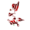
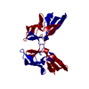
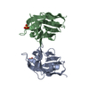
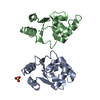
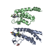
- PDB-4j4c: Structure of P51G Cyanovirin-N swapped dimer in the P3221 space group -
+ Open data

- Basic information
| Entry | Database: PDB / ID: 4j4c | ||||||
|---|---|---|---|---|---|---|---|
| Title | Structure of P51G Cyanovirin-N swapped dimer in the P3221 space group | ||||||
 Components Components | Cyanovirin-N | ||||||
 Keywords Keywords | SUGAR BINDING PROTEIN / CVNH fold / carbohydrate binding protein / antiviral protein | ||||||
| Function / homology |  Function and homology information Function and homology informationoligosaccharide binding / regulation of defense response to virus / carbohydrate binding Similarity search - Function | ||||||
| Biological species |  Nostoc ellipsosporum (bacteria) Nostoc ellipsosporum (bacteria) | ||||||
| Method |  X-RAY DIFFRACTION / MOLECULAR REPLACEMENT / Resolution: 1.9 Å X-RAY DIFFRACTION / MOLECULAR REPLACEMENT / Resolution: 1.9 Å | ||||||
 Authors Authors | Koharudin, L.M.I. / Liu, L. / Gronenborn, A.M. | ||||||
 Citation Citation | Journal: Proc.Natl.Acad.Sci.USA / Year: 2013 Title: Different 3D domain-swapped oligomeric cyanovirin-N structures suggest trapped folding intermediates. Authors: Koharudin, L.M. / Liu, L. / Gronenborn, A.M. | ||||||
| History |
|
- Structure visualization
| Structure viewer | Molecule: MolmilJmol/JSmol |
|---|
- Downloads & links
-Download
| PDBx/mmCIF format | 4j4c.cif.gz | 34.5 KB | Display | PDBx/mmCIF format |
|---|---|---|---|---|
| PDB format | pdb4j4c.ent.gz | 23.3 KB | Display | PDB format |
| PDBx/mmJSON format | 4j4c.json.gz | Tree view | PDBx/mmJSON format | |
| Others |  Other downloads Other downloads |
-Validation report
| Arichive directory | https://data.pdbj.org/pub/pdb/validation_reports/j4/4j4cftp://data.pdbj.org/pub/pdb/validation_reports/j4/4j4c | HTTPS FTP |
|---|
-Related structure data
| Related structure data |  4j4dC  4j4eC  4j4fC  4j4gC  3ezmS C: citing same article ( S: Starting model for refinement |
|---|---|
| Similar structure data |

-Links
 PDBj
PDBj- Assembly
| Deposited unit | 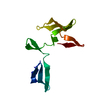
| |||||||||
|---|---|---|---|---|---|---|---|---|---|---|
| 1 | 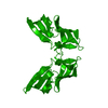
| |||||||||
| Unit cell |
| |||||||||
| Components on special symmetry positions |
|
-Components
| #1: Protein | Mass: 10982.026 Da / Num. of mol.: 1 / Mutation: P51G Source method: isolated from a genetically manipulated source Source: (gene. exp.) Nostoc ellipsosporum (bacteria) / Plasmid: pET26B / Production host: |
|---|---|
| #2: Water | ChemComp-HOH /  Mass: 18.015 Da / Num. of mol.: 86 / Source method: isolated from a natural source / Formula: H2O Mass: 18.015 Da / Num. of mol.: 86 / Source method: isolated from a natural source / Formula: H2O |
| Has protein modification | Y |
-Experimental details
-Experiment
| Experiment | Method: X-RAY DIFFRACTION / Number of used crystals: 1 |
|---|
- Sample preparation
| Crystal | Density Matthews: 2.38 Å3/Da / Density % sol: 48.31 % |
|---|---|
| Crystal grow | Temperature: 295 K / Method: vapor diffusion, sitting drop / pH: 6 Details: 20% w/v PEG8000, 0.1 M sodium phosphate/citrate, pH 4.2, 0.2 M sodium chloride, VAPOR DIFFUSION, SITTING DROP, temperature 295K |
-Data collection
| Diffraction | Mean temperature: 93 K |
|---|---|
| Diffraction source | Source: ROTATING ANODE / Type: RIGAKU FR-E SUPERBRIGHT / Wavelength: 1.5418 Å |
| Detector | Type: RIGAKU / Detector: IMAGE PLATE / Date: Nov 12, 2011 / Details: HF VariMax |
| Radiation | Monochromator: OSMIC MIRRORS / Protocol: SINGLE WAVELENGTH / Monochromatic (M) / Laue (L): M / Scattering type: x-ray |
| Radiation wavelength | Wavelength: 1.5418 Å / Relative weight: 1 |
| Reflection | Resolution: 1.9→36.73 Å / Num. all: 8694 / Num. obs: 8659 / % possible obs: 99.6 % / Observed criterion σ(F): 3 / Redundancy: 14.28 % / Rmerge(I) obs: 0.08 / Net I/σ(I): 18.6 |
| Reflection shell | Resolution: 1.9→1.97 Å / Redundancy: 14.17 % / Rmerge(I) obs: 0.433 / Mean I/σ(I) obs: 5 / Num. unique all: 612 / % possible all: 98.1 |
- Processing
| Software |
| |||||||||||||||||||||||||||||||||||||||||||||||||||||||||||||||||
|---|---|---|---|---|---|---|---|---|---|---|---|---|---|---|---|---|---|---|---|---|---|---|---|---|---|---|---|---|---|---|---|---|---|---|---|---|---|---|---|---|---|---|---|---|---|---|---|---|---|---|---|---|---|---|---|---|---|---|---|---|---|---|---|---|---|---|
| Refinement | Method to determine structure: MOLECULAR REPLACEMENT Starting model: PDB ENTRY 3EZM Resolution: 1.9→36.73 Å / Cor.coef. Fo:Fc: 0.955 / Cor.coef. Fo:Fc free: 0.934 / SU B: 3.286 / SU ML: 0.096 / Isotropic thermal model: ISOTROPIC / Cross valid method: THROUGHOUT / σ(F): 1 / ESU R: 0.159 / ESU R Free: 0.142 / Stereochemistry target values: MAXIMUM LIKELIHOOD Details: HYDROGENS HAVE BEEN ADDED IN THE RIDING POSITIONS U VALUES : REFINED INDIVIDUALLY
| |||||||||||||||||||||||||||||||||||||||||||||||||||||||||||||||||
| Solvent computation | Ion probe radii: 0.8 Å / Shrinkage radii: 0.8 Å / VDW probe radii: 1.4 Å / Solvent model: BABINET MODEL WITH MASK | |||||||||||||||||||||||||||||||||||||||||||||||||||||||||||||||||
| Displacement parameters | Biso mean: 46.744 Å2
| |||||||||||||||||||||||||||||||||||||||||||||||||||||||||||||||||
| Refinement step | Cycle: LAST / Resolution: 1.9→36.73 Å
| |||||||||||||||||||||||||||||||||||||||||||||||||||||||||||||||||
| Refine LS restraints |
| |||||||||||||||||||||||||||||||||||||||||||||||||||||||||||||||||
| LS refinement shell | Resolution: 1.9→1.948 Å / Total num. of bins used: 20
|
