 Movie
Movie Controller

[English] 日本語
 Yorodumi
- PDB-4j16: Crystal structure of Thermus thermophilus transhydrogenase hetero... -
+ Open data

- Basic information
| Entry | Database: PDB / ID: 4j16 | ||||||
|---|---|---|---|---|---|---|---|
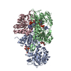
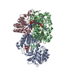
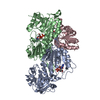
| Title | Crystal structure of Thermus thermophilus transhydrogenase heterotrimeric complex of the Alpha1 subunit dimer with the NADP binding domain (domain III) of the Beta subunit | ||||||
 Components Components |
| ||||||
 Keywords Keywords | OXIDOREDUCTASE / Soluble components of nicotinamide nucleotide transhydrogenase / Complex of Alpha1 subunit dimer with Domain III of Beta subunit / Alpha1 binds NAD(H) / Domain III binds NADP(H) / Domain III binds to Alpha1 / NAD bound to Alpha1 / NADP bound to Domain III | ||||||
| Function / homology |  Function and homology information Function and homology informationproton-translocating NAD(P)+ transhydrogenase activity / proton-translocating NAD(P)+ transhydrogenase / NADPH regeneration / NADP binding / oxidoreductase activity / plasma membrane Similarity search - Function | ||||||
| Biological species |   Thermus thermophilus (bacteria) Thermus thermophilus (bacteria) | ||||||
| Method |  X-RAY DIFFRACTION / SYNCHROTRON / MOLECULAR REPLACEMENT / Resolution: 2.41 Å X-RAY DIFFRACTION / SYNCHROTRON / MOLECULAR REPLACEMENT / Resolution: 2.41 Å | ||||||
 Authors Authors | Yamaguchi, M. / Leung, J. / Schurig Briccio, L.A. / Gennis, R.B. / Stout, C.D. | ||||||
 Citation Citation | Journal: To be Published Title: Crystal structure analysis of Thermus thermophilus transhydrogenase soluble domains Authors: Yamaguchi, M. / Leung, J. / Schurig Briccio, L.A. / Gennis, R.B. / Stout, C.D. | ||||||
| History |
|
- Structure visualization
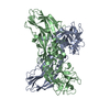
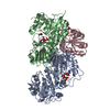
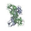
| Structure viewer | Molecule: MolmilJmol/JSmol |
|---|
- Downloads & links
-Download
| PDBx/mmCIF format | 4j16.cif.gz | 368.6 KB | Display | PDBx/mmCIF format |
|---|---|---|---|---|
| PDB format | pdb4j16.ent.gz | 301.7 KB | Display | PDB format |
| PDBx/mmJSON format | 4j16.json.gz | Tree view | PDBx/mmJSON format | |
| Others |  Other downloads Other downloads |
-Validation report
| Arichive directory | https://data.pdbj.org/pub/pdb/validation_reports/j1/4j16ftp://data.pdbj.org/pub/pdb/validation_reports/j1/4j16 | HTTPS FTP |
|---|
-Related structure data
| Related structure data | 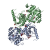 4izhSC 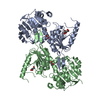 4iziC 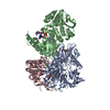 4j1tC  1pnoS C: citing same article ( S: Starting model for refinement |
|---|---|
| Similar structure data |


-Links
 PDBj
PDBj

- Assembly
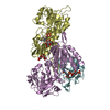
| Deposited unit | 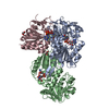
| ||||||||
|---|---|---|---|---|---|---|---|---|---|
| 1 |
| ||||||||
| Unit cell |
|
-Components
-Protein , 2 types, 3 molecules ABC
| #1: Protein | Mass: 40851.340 Da / Num. of mol.: 2 Source method: isolated from a genetically manipulated source Source: (gene. exp.) Thermus thermophilus (bacteria) / Strain: HB27 / ATCC BAA-163 / DSM 7039 / Gene: TT_C1780 / Plasmid: pet21a / Production host: #2: Protein | | Mass: 20018.150 Da / Num. of mol.: 1 / Fragment: Domain III (UNP residues 266-450) Source method: isolated from a genetically manipulated source Source: (gene. exp.) Thermus thermophilus (bacteria) / Strain: HB27 / ATCC BAA-163 / DSM 7039 / Plasmid: pet21a / Production host: |
|---|
-Non-polymers , 6 types, 128 molecules 


| #3: Chemical |  Mass: 663.425 Da / Num. of mol.: 2 / Source method: obtained synthetically / Formula: C21H27N7O14P2 / Comment: NAD*YM Mass: 663.425 Da / Num. of mol.: 2 / Source method: obtained synthetically / Formula: C21H27N7O14P2 / Comment: NAD*YM#4: Chemical | ChemComp-CL / |  Mass: 35.453 Da / Num. of mol.: 1 / Source method: obtained synthetically / Formula: Cl Mass: 35.453 Da / Num. of mol.: 1 / Source method: obtained synthetically / Formula: Cl#5: Chemical | ChemComp-PGE / |  Mass: 150.173 Da / Num. of mol.: 1 / Source method: obtained synthetically / Formula: C6H14O4 Mass: 150.173 Da / Num. of mol.: 1 / Source method: obtained synthetically / Formula: C6H14O4#6: Chemical |  Mass: 92.094 Da / Num. of mol.: 2 / Source method: obtained synthetically / Formula: C3H8O3 Mass: 92.094 Da / Num. of mol.: 2 / Source method: obtained synthetically / Formula: C3H8O3#7: Chemical | ChemComp-NAP / |  Mass: 743.405 Da / Num. of mol.: 1 / Source method: obtained synthetically / Formula: C21H28N7O17P3 Mass: 743.405 Da / Num. of mol.: 1 / Source method: obtained synthetically / Formula: C21H28N7O17P3#8: Water | ChemComp-HOH / | Mass: 18.015 Da / Num. of mol.: 121 / Source method: isolated from a natural source / Formula: H2O |
|---|
-Experimental details
-Experiment
| Experiment | Method: X-RAY DIFFRACTION / Number of used crystals: 1 |
|---|
- Sample preparation
| Crystal | Density Matthews: 2.47 Å3/Da / Density % sol: 50.22 % |
|---|---|
| Crystal grow | Temperature: 291 K / Method: vapor diffusion, sitting drop / pH: 6.5 Details: Molecular Dimensions MD1-47 Morpheus kit, condition E2: 0.12 M ethylene glycols, 0.1 M pH 6.5 buffers, 30% ethylene glycol + PEG8000, VAPOR DIFFUSION, SITTING DROP, temperature 291K |
-Data collection
| Diffraction | Mean temperature: 100 K |
|---|---|
| Diffraction source | Source: SYNCHROTRON / Site: SSRL  / Beamline: BL7-1 / Wavelength: 1.18076 Å / Beamline: BL7-1 / Wavelength: 1.18076 Å |
| Detector | Type: ADSC QUANTUM 315r / Detector: CCD / Date: Dec 2, 2011 / Details: Rh coated flat mirror |
| Radiation | Monochromator: Side scattering I-beam bent single crystal, asymmetric cut 4.9650 degrees Protocol: SINGLE WAVELENGTH / Monochromatic (M) / Laue (L): M / Scattering type: x-ray |
| Radiation wavelength | Wavelength: 1.18076 Å / Relative weight: 1 |
| Reflection | Resolution: 2.4→39.98 Å / Num. all: 38049 / Num. obs: 36638 / % possible obs: 96.29 % / Observed criterion σ(F): 0 / Observed criterion σ(I): 0 / Redundancy: 3.7 % / Biso Wilson estimate: 62 Å2 / Rmerge(I) obs: 0.036 / Rsym value: 0.036 / Net I/σ(I): 18.4 |
| Reflection shell | Resolution: 2.4→2.53 Å / Redundancy: 2.6 % / Rmerge(I) obs: 0.557 / Mean I/σ(I) obs: 1.4 / Num. unique all: 4538 / Rsym value: 0.557 / % possible all: 79.1 |
- Processing
| Software |
| ||||||||||||||||||||||||||||||||||||||||||||||||||||||||||||||||||||||||||||||||||||||||||||||||||||
|---|---|---|---|---|---|---|---|---|---|---|---|---|---|---|---|---|---|---|---|---|---|---|---|---|---|---|---|---|---|---|---|---|---|---|---|---|---|---|---|---|---|---|---|---|---|---|---|---|---|---|---|---|---|---|---|---|---|---|---|---|---|---|---|---|---|---|---|---|---|---|---|---|---|---|---|---|---|---|---|---|---|---|---|---|---|---|---|---|---|---|---|---|---|---|---|---|---|---|---|---|---|
| Refinement | Method to determine structure: MOLECULAR REPLACEMENT Starting model: PDB ENTRIES 4IZH, 1PNO Resolution: 2.41→39.98 Å / Cor.coef. Fo:Fc: 0.956 / Cor.coef. Fo:Fc free: 0.926 / SU B: 20.836 / SU ML: 0.227 / Cross valid method: THROUGHOUT / σ(F): 0 / σ(I): 0 / ESU R: 0.474 / ESU R Free: 0.278 / Stereochemistry target values: MAXIMUM LIKELIHOOD / Details: HYDROGENS HAVE BEEN ADDED IN THE RIDING POSITIONS
| ||||||||||||||||||||||||||||||||||||||||||||||||||||||||||||||||||||||||||||||||||||||||||||||||||||
| Solvent computation | Ion probe radii: 0.8 Å / Shrinkage radii: 0.8 Å / VDW probe radii: 1.2 Å / Solvent model: MASK | ||||||||||||||||||||||||||||||||||||||||||||||||||||||||||||||||||||||||||||||||||||||||||||||||||||
| Displacement parameters | Biso mean: 73.164 Å2
| ||||||||||||||||||||||||||||||||||||||||||||||||||||||||||||||||||||||||||||||||||||||||||||||||||||
| Refinement step | Cycle: LAST / Resolution: 2.41→39.98 Å
| ||||||||||||||||||||||||||||||||||||||||||||||||||||||||||||||||||||||||||||||||||||||||||||||||||||
| Refine LS restraints |
| ||||||||||||||||||||||||||||||||||||||||||||||||||||||||||||||||||||||||||||||||||||||||||||||||||||
| LS refinement shell | Resolution: 2.41→2.467 Å / Total num. of bins used: 20
| ||||||||||||||||||||||||||||||||||||||||||||||||||||||||||||||||||||||||||||||||||||||||||||||||||||
| Refinement TLS params. | Method: refined / Refine-ID: X-RAY DIFFRACTION
| ||||||||||||||||||||||||||||||||||||||||||||||||||||||||||||||||||||||||||||||||||||||||||||||||||||
| Refinement TLS group |
|
