 Movie
Movie Controller

[English] 日本語
 Yorodumi
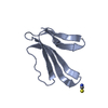
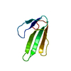
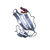
- PDB-4iye: Crystal structure of AdTx1 (rho-Da1a) from eastern green mamba (D... -
+ Open data

- Basic information
| Entry | Database: PDB / ID: 4iye | ||||||
|---|---|---|---|---|---|---|---|
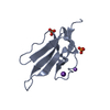
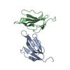
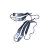
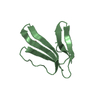
| Title | Crystal structure of AdTx1 (rho-Da1a) from eastern green mamba (Dendroaspis angusticeps) | ||||||
 Components Components | Toxin AdTx1 | ||||||
 Keywords Keywords | TOXIN / Snake three-finger toxin family / Type A muscarinic toxin subfamily / Allosteric antagonist of the alpha-1A adrenergic receptor (ADRA1A) / Acts as a relaxant of smooth muscle / alpha-1A adrenergic receptor / g-rhoDa1a K34A / Expressed by the venom gland | ||||||
| Function / homology |  Function and homology information Function and homology information | ||||||
| Biological species |  Dendroaspis angusticeps (eastern green mamba) Dendroaspis angusticeps (eastern green mamba) | ||||||
| Method |  X-RAY DIFFRACTION / SYNCHROTRON / MOLECULAR REPLACEMENT / Resolution: 1.951 Å X-RAY DIFFRACTION / SYNCHROTRON / MOLECULAR REPLACEMENT / Resolution: 1.951 Å | ||||||
 Authors Authors | Stura, E.A. / Vera, L. / Maiga, A.A. / Marchetti, C. / Lorphelin, A. / Bellanger, L. / Servant, D. / Gilles, N. | ||||||
 Citation Citation | Journal: Acta Crystallogr.,Sect.F / Year: 2013 Title: Crystallization of recombinant green mamba rho-Da1a toxin during a lyophilization procedure and its structure determination. Authors: Maiga, A. / Vera, L. / Marchetti, C. / Lorphelin, A. / Bellanger, L. / Mourier, G. / Servent, D. / Gilles, N. / Stura, E.A. #1: Journal: Acta Crystallogr.,Sect.FTitle: Crystallization of Da1a from Green mamba venom during lyophilization | ||||||
| History |
|
- Structure visualization
| Structure viewer | Molecule: MolmilJmol/JSmol |
|---|
- Downloads & links
-Download
| PDBx/mmCIF format | 4iye.cif.gz | 41.6 KB | Display | PDBx/mmCIF format |
|---|---|---|---|---|
| PDB format | pdb4iye.ent.gz | 28.3 KB | Display | PDB format |
| PDBx/mmJSON format | 4iye.json.gz | Tree view | PDBx/mmJSON format | |
| Others |  Other downloads Other downloads |
-Validation report
| Arichive directory | https://data.pdbj.org/pub/pdb/validation_reports/iy/4iyeftp://data.pdbj.org/pub/pdb/validation_reports/iy/4iye | HTTPS FTP |
|---|
-Related structure data
| Related structure data |  1ff4S S: Starting model for refinement |
|---|---|
| Similar structure data |


-Links
 PDBj
PDBj

- Assembly
| Deposited unit | 
| ||||||||
|---|---|---|---|---|---|---|---|---|---|
| 1 |
| ||||||||
| Unit cell |
|
-Components
| #1: Protein | Mass: 7300.272 Da / Num. of mol.: 1 / Fragment: Rho-Da1a / Mutation: K34A Source method: isolated from a genetically manipulated source Source: (gene. exp.) Dendroaspis angusticeps (eastern green mamba)Strain: angusticeps / Organ: Venom gland Plasmid details: The synthetic gene corresponds to the ENLYFQG-rho-Da1a protein flanked by the attB1 and attB2 sequences Plasmid: pENTRE plasmid / Production host:  | ||||
|---|---|---|---|---|---|
| #2: Chemical | ChemComp-EDO /   Mass: 62.068 Da / Num. of mol.: 1 / Source method: obtained synthetically / Formula: C2H6O2 Mass: 62.068 Da / Num. of mol.: 1 / Source method: obtained synthetically / Formula: C2H6O2 | ||||
| #3: Chemical |   Mass: 106.120 Da / Num. of mol.: 2 / Source method: obtained synthetically / Formula: C4H10O3 Mass: 106.120 Da / Num. of mol.: 2 / Source method: obtained synthetically / Formula: C4H10O3#4: Water | ChemComp-HOH / |  Mass: 18.015 Da / Num. of mol.: 70 / Source method: isolated from a natural source / Formula: H2O Mass: 18.015 Da / Num. of mol.: 70 / Source method: isolated from a natural source / Formula: H2OHas protein modification | Y | |
-Experimental details
-Experiment
| Experiment | Method: X-RAY DIFFRACTION / Number of used crystals: 1 |
|---|
- Sample preparation
| Crystal | Density Matthews: 1.82 Å3/Da / Density % sol: 32.56 % |
|---|---|
| Crystal grow | Temperature: 273 K / Method: lyophilization / pH: 8 Details: Crystallization during lyophilization due to increased concentration and low temperature. Cryoconditions: 27% PEG8K, 15% MPEG550, 10% glycerol, 0.09 M Tris-HCl, pH 8.0, Lyophilization, temperature 273K |
-Data collection
| Diffraction | Mean temperature: 100 K | ||||||||||||||||||||||||||||||||||||||||||||||||||||||||||||||||||||||
|---|---|---|---|---|---|---|---|---|---|---|---|---|---|---|---|---|---|---|---|---|---|---|---|---|---|---|---|---|---|---|---|---|---|---|---|---|---|---|---|---|---|---|---|---|---|---|---|---|---|---|---|---|---|---|---|---|---|---|---|---|---|---|---|---|---|---|---|---|---|---|---|
| Diffraction source | Source: SYNCHROTRON / Site: ESRF  / Beamline: ID14-1 / Wavelength: 0.9334 Å / Beamline: ID14-1 / Wavelength: 0.9334 Å | ||||||||||||||||||||||||||||||||||||||||||||||||||||||||||||||||||||||
| Detector | Type: ADSC QUANTUM 4 / Detector: CCD / Date: Jun 21, 2010 / Details: mirrors | ||||||||||||||||||||||||||||||||||||||||||||||||||||||||||||||||||||||
| Radiation | Monochromator: Si 111 CHANNEL / Protocol: SINGLE WAVELENGTH / Monochromatic (M) / Laue (L): M / Scattering type: x-ray | ||||||||||||||||||||||||||||||||||||||||||||||||||||||||||||||||||||||
| Radiation wavelength | Wavelength: 0.9334 Å / Relative weight: 1 | ||||||||||||||||||||||||||||||||||||||||||||||||||||||||||||||||||||||
| Reflection | Resolution: 1.95→32.36 Å / Num. all: 4193 / Num. obs: 3937 / % possible obs: 93.9 % / Observed criterion σ(F): 0 / Observed criterion σ(I): -4 / Redundancy: 8.44 % / Biso Wilson estimate: 25.325 Å2 / Rmerge(I) obs: 0.171 / Rsym value: 0.16 / Net I/σ(I): 11.43 | ||||||||||||||||||||||||||||||||||||||||||||||||||||||||||||||||||||||
| Reflection shell | Diffraction-ID: 1
|
- Processing
| Software |
| |||||||||||||||||||||||||||||||||||||||||||||||||||||||||||||||||||||||||||||||||||||||||||||||||||||||||||||||||||||||||||||||||||||||||||||||||||
|---|---|---|---|---|---|---|---|---|---|---|---|---|---|---|---|---|---|---|---|---|---|---|---|---|---|---|---|---|---|---|---|---|---|---|---|---|---|---|---|---|---|---|---|---|---|---|---|---|---|---|---|---|---|---|---|---|---|---|---|---|---|---|---|---|---|---|---|---|---|---|---|---|---|---|---|---|---|---|---|---|---|---|---|---|---|---|---|---|---|---|---|---|---|---|---|---|---|---|---|---|---|---|---|---|---|---|---|---|---|---|---|---|---|---|---|---|---|---|---|---|---|---|---|---|---|---|---|---|---|---|---|---|---|---|---|---|---|---|---|---|---|---|---|---|---|---|---|---|
| Refinement | Method to determine structure: MOLECULAR REPLACEMENT Starting model: PDB ENTRY 1FF4 without loop 1 tip Resolution: 1.951→32.36 Å / Cor.coef. Fo:Fc: 0.959 / Cor.coef. Fo:Fc free: 0.899 / SU B: 12.031 / SU ML: 0.156 / Isotropic thermal model: Anisotropic TLS / Cross valid method: THROUGHOUT / σ(F): 0 / σ(I): -3 / ESU R Free: 0.204 / Stereochemistry target values: MAXIMUM LIKELIHOOD / Details: HYDROGENS HAVE BEEN ADDED IN THE RIDING POSITIONS
| |||||||||||||||||||||||||||||||||||||||||||||||||||||||||||||||||||||||||||||||||||||||||||||||||||||||||||||||||||||||||||||||||||||||||||||||||||
| Solvent computation | Ion probe radii: 0.8 Å / Shrinkage radii: 0.8 Å / VDW probe radii: 1.2 Å / Solvent model: MASK | |||||||||||||||||||||||||||||||||||||||||||||||||||||||||||||||||||||||||||||||||||||||||||||||||||||||||||||||||||||||||||||||||||||||||||||||||||
| Displacement parameters | Biso mean: 25.704 Å2
| |||||||||||||||||||||||||||||||||||||||||||||||||||||||||||||||||||||||||||||||||||||||||||||||||||||||||||||||||||||||||||||||||||||||||||||||||||
| Refinement step | Cycle: LAST / Resolution: 1.951→32.36 Å
| |||||||||||||||||||||||||||||||||||||||||||||||||||||||||||||||||||||||||||||||||||||||||||||||||||||||||||||||||||||||||||||||||||||||||||||||||||
| Refine LS restraints |
| |||||||||||||||||||||||||||||||||||||||||||||||||||||||||||||||||||||||||||||||||||||||||||||||||||||||||||||||||||||||||||||||||||||||||||||||||||
| LS refinement shell | Refine-ID: X-RAY DIFFRACTION / Total num. of bins used: 20
| |||||||||||||||||||||||||||||||||||||||||||||||||||||||||||||||||||||||||||||||||||||||||||||||||||||||||||||||||||||||||||||||||||||||||||||||||||
| Refinement TLS params. | Method: refined / Origin x: -16.1157 Å / Origin y: 12.5129 Å / Origin z: 13.2222 Å
|
