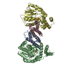
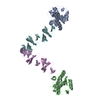
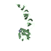
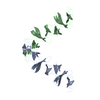
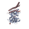
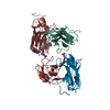
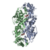
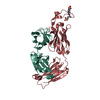
Journal: mBio / Year: 2014 Title: Structural and functional insights into peptidoglycan access for the lytic amidase LytA of Streptococcus pneumoniae. Abstract: The cytosolic N-acetylmuramoyl-l-alanine amidase LytA protein of Streptococcus pneumoniae, which is released by bacterial lysis, associates with the cell wall via its choline-binding motif. During ...The cytosolic N-acetylmuramoyl-l-alanine amidase LytA protein of Streptococcus pneumoniae, which is released by bacterial lysis, associates with the cell wall via its choline-binding motif. During exponential growth, LytA accesses its peptidoglycan substrate to cause lysis only when nascent peptidoglycan synthesis is stalled by nutrient starvation or β-lactam antibiotics. Here we present three-dimensional structures of LytA and establish the requirements for substrate binding and catalytic activity. The solution structure of the full-length LytA dimer reveals a peculiar fold, with the choline-binding domains forming a rigid V-shaped scaffold and the relatively more flexible amidase domains attached in a trans position. The 1.05-Å crystal structure of the amidase domain reveals a prominent Y-shaped binding crevice composed of three contiguous subregions, with a zinc-containing active site localized at the bottom of the branch point. Site-directed mutagenesis was employed to identify catalytic residues and to investigate the relative impact of potential substrate-interacting residues lining the binding crevice for the lytic activity of LytA. In vitro activity assays using defined muropeptide substrates reveal that LytA utilizes a large substrate recognition interface and requires large muropeptide substrates with several connected saccharides that interact with all subregions of the binding crevice for catalysis. We hypothesize that the substrate requirements restrict LytA to the sites on the cell wall where nascent peptidoglycan synthesis occurs. IMPORTANCE: Streptococcus pneumoniae is a human respiratory tract pathogen responsible for millions of deaths annually. Its major pneumococcal autolysin, LytA, is required for autolysis and ...IMPORTANCE: Streptococcus pneumoniae is a human respiratory tract pathogen responsible for millions of deaths annually. Its major pneumococcal autolysin, LytA, is required for autolysis and fratricidal lysis and functions as a virulence factor that facilitates the spread of toxins and factors involved in immune evasion. LytA is also activated by penicillin and vancomycin and is responsible for the lysis induced by these antibiotics. The factors that regulate the lytic activity of LytA are unclear, but it was recently demonstrated that control is at the level of substrate recognition and that LytA required access to the nascent peptidoglycan. The present study was undertaken to structurally and functionally investigate LytA and its substrate-interacting interface and to determine the requirements for substrate recognition and catalysis. Our results reveal that the amidase domain comprises a complex substrate-binding crevice and needs to interact with a large-motif epitope of peptidoglycan for catalysis.
Resolution: 2.6→30 Å / Cor.coef. Fo:Fc: 0.939 / Cor.coef. Fo:Fc free: 0.878 / SU B: 12.946 / SU ML: 0.268 / Cross valid method: THROUGHOUT / ESU R: 4.141 / ESU R Free: 0.368 / Stereochemistry target values: MAXIMUM LIKELIHOOD / Details: HYDROGENS HAVE BEEN USED IF PRESENT IN THE INPUT
Rfactor
Num. reflection
% reflection
Selection details
Rfree
0.27337
511
4.8 %
RANDOM
Rwork
0.19836
-
-
-
obs
0.20176
10078
93.72 %
-
all
-
10078
-
-
Solvent computation
Ion probe radii: 0.8 Å / Shrinkage radii: 0.8 Å / VDW probe radii: 1.2 Å / Solvent model: MASK
Displacement parameters
Biso mean: 35.195 Å2
Baniso -1
Baniso -2
Baniso -3
1-
3.54 Å2
-0 Å2
-0.78 Å2
2-
-
-4.74 Å2
0 Å2
3-
-
-
1.24 Å2
Refinement step
Cycle: LAST / Resolution: 2.6→30 Å
Protein
Nucleic acid
Ligand
Solvent
Total
Num. atoms
2408
0
70
31
2509
Refine LS restraints
Refine-ID
Type
Dev ideal
Dev ideal target
Number
X-RAY DIFFRACTION
r_bond_refined_d
0.012
0.02
2560
X-RAY DIFFRACTION
r_bond_other_d
X-RAY DIFFRACTION
r_angle_refined_deg
1.714
1.932
3474
X-RAY DIFFRACTION
r_angle_other_deg
X-RAY DIFFRACTION
r_dihedral_angle_1_deg
8.763
5
290
X-RAY DIFFRACTION
r_dihedral_angle_2_deg
33.207
24.262
122
X-RAY DIFFRACTION
r_dihedral_angle_3_deg
22.396
15
382
X-RAY DIFFRACTION
r_dihedral_angle_4_deg
9.743
15
2
X-RAY DIFFRACTION
r_chiral_restr
0.143
0.2
320
X-RAY DIFFRACTION
r_gen_planes_refined
0.007
0.02
1974
X-RAY DIFFRACTION
r_gen_planes_other
X-RAY DIFFRACTION
r_nbd_refined
X-RAY DIFFRACTION
r_nbd_other
X-RAY DIFFRACTION
r_nbtor_refined
X-RAY DIFFRACTION
r_nbtor_other
X-RAY DIFFRACTION
r_xyhbond_nbd_refined
X-RAY DIFFRACTION
r_xyhbond_nbd_other
X-RAY DIFFRACTION
r_metal_ion_refined
X-RAY DIFFRACTION
r_metal_ion_other
X-RAY DIFFRACTION
r_symmetry_vdw_refined
X-RAY DIFFRACTION
r_symmetry_vdw_other
X-RAY DIFFRACTION
r_symmetry_hbond_refined
X-RAY DIFFRACTION
r_symmetry_hbond_other
X-RAY DIFFRACTION
r_symmetry_metal_ion_refined
X-RAY DIFFRACTION
r_symmetry_metal_ion_other
X-RAY DIFFRACTION
r_mcbond_it
X-RAY DIFFRACTION
r_mcbond_other
X-RAY DIFFRACTION
r_mcangle_it
X-RAY DIFFRACTION
r_scbond_it
X-RAY DIFFRACTION
r_scangle_it
X-RAY DIFFRACTION
r_rigid_bond_restr
X-RAY DIFFRACTION
r_sphericity_free
X-RAY DIFFRACTION
r_sphericity_bonded
LS refinement shell
Resolution: 2.6→2.667 Å / Total num. of bins used: 20
Rfactor
Num. reflection
% reflection
Rfree
0.409
34
-
Rwork
0.341
709
-
obs
-
-
89.41 %
+
About Yorodumi
-
News
-
Feb 9, 2022. New format data for meta-information of EMDB entries
New format data for meta-information of EMDB entries
Version 3 of the EMDB header file is now the official format.
The previous official version 1.9 will be removed from the archive.
In the structure databanks used in Yorodumi, some data are registered as the other names, "COVID-19 virus" and "2019-nCoV". Here are the details of the virus and the list of structure data.
Jan 31, 2019. EMDB accession codes are about to change! (news from PDBe EMDB page)
EMDB accession codes are about to change! (news from PDBe EMDB page)
The allocation of 4 digits for EMDB accession codes will soon come to an end. Whilst these codes will remain in use, new EMDB accession codes will include an additional digit and will expand incrementally as the available range of codes is exhausted. The current 4-digit format prefixed with “EMD-” (i.e. EMD-XXXX) will advance to a 5-digit format (i.e. EMD-XXXXX), and so on. It is currently estimated that the 4-digit codes will be depleted around Spring 2019, at which point the 5-digit format will come into force.
The EM Navigator/Yorodumi systems omit the EMD- prefix.
Related info.:Q: What is EMD? / ID/Accession-code notation in Yorodumi/EM Navigator
Yorodumi is a browser for structure data from EMDB, PDB, SASBDB, etc.
This page is also the successor to EM Navigator detail page, and also detail information page/front-end page for Omokage search.
The word "yorodu" (or yorozu) is an old Japanese word meaning "ten thousand". "mi" (miru) is to see.
Related info.:EMDB / PDB / SASBDB / Comparison of 3 databanks / Yorodumi Search / Aug 31, 2016. New EM Navigator & Yorodumi / Yorodumi Papers / Jmol/JSmol / Function and homology information / Changes in new EM Navigator and Yorodumi
 Movie
Movie Controller

 Yorodumi
 Open data
 Basic information
 Components
 Keywords
 Function and homology information
 Streptococcus pneumoniae (bacteria)
 X-RAY DIFFRACTION /
  Authors
 Citation
 Structure visualization
 Downloads & links
 Other downloads


 PDBj
PDBj Assembly
 Mass: 104.171 Da / Num. of mol.: 10 / Source method: obtained synthetically / Formula: C5H14NO
 Mass: 18.015 Da / Num. of mol.: 31 / Source method: isolated from a natural source / Formula: H2O
 Sample preparation
 / Beamline: ID29 / Wavelength: 0.9762 Å
 Processing