 Movie
Movie Controller

[English] 日本語
 Yorodumi
- PDB-4iuk: Crystal structure of NreA of Staphylococcus carnosus with bound n... -
+ Open data

- Basic information
| Entry | Database: PDB / ID: 4iuk | ||||||
|---|---|---|---|---|---|---|---|
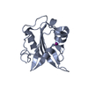
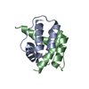
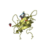
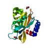
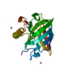
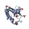
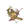
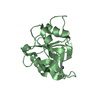
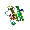
| Title | Crystal structure of NreA of Staphylococcus carnosus with bound nitrate | ||||||
 Components Components | NreA protein | ||||||
 Keywords Keywords | Nitrate-binding protein / GAF domain / Nitrate sensor / Staphylococcus / Nitrate binding / Nitrate / Cytosolic | ||||||
| Function / homology | GAF domain / GAF domain / GAF domain / GAF-like domain superfamily / Beta-Lactamase / 2-Layer Sandwich / Alpha Beta / NITRATE ION / NreA protein Function and homology information Function and homology information | ||||||
| Biological species |  Staphylococcus carnosus subsp. carnosus (bacteria) Staphylococcus carnosus subsp. carnosus (bacteria) | ||||||
| Method |  X-RAY DIFFRACTION / SYNCHROTRON / MOLECULAR REPLACEMENT / Resolution: 2.35 Å X-RAY DIFFRACTION / SYNCHROTRON / MOLECULAR REPLACEMENT / Resolution: 2.35 Å | ||||||
 Authors Authors | Stehle, T. / Niemann, V. | ||||||
 Citation Citation | Journal: J.Mol.Biol. / Year: 2014 Title: The NreA Protein Functions as a Nitrate Receptor in the Staphylococcal Nitrate Regulation System. Authors: Niemann, V. / Koch-Singenstreu, M. / Neu, A. / Nilkens, S. / Gotz, F. / Unden, G. / Stehle, T. | ||||||
| History |
|
- Structure visualization
| Structure viewer | Molecule: MolmilJmol/JSmol |
|---|
- Downloads & links
-Download
| PDBx/mmCIF format | 4iuk.cif.gz | 122.8 KB | Display | PDBx/mmCIF format |
|---|---|---|---|---|
| PDB format | pdb4iuk.ent.gz | 95.8 KB | Display | PDB format |
| PDBx/mmJSON format | 4iuk.json.gz | Tree view | PDBx/mmJSON format | |
| Others |  Other downloads Other downloads |
-Validation report
| Arichive directory | https://data.pdbj.org/pub/pdb/validation_reports/iu/4iukftp://data.pdbj.org/pub/pdb/validation_reports/iu/4iuk | HTTPS FTP |
|---|
-Related structure data


-Links
 PDBj
PDBj

- Assembly
| Deposited unit | 
| ||||||||
|---|---|---|---|---|---|---|---|---|---|
| 1 | 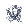
| ||||||||
| 2 | 
| ||||||||
| Unit cell |
| ||||||||
| Components on special symmetry positions |
|
-Components
| #1: Protein | Mass: 18854.338 Da / Num. of mol.: 2 Source method: isolated from a genetically manipulated source Source: (gene. exp.) Staphylococcus carnosus subsp. carnosus (bacteria)Strain: TM300 / Gene: nreA, Sca_1890, SCA_1890 / Production host: #2: Chemical |   Mass: 62.005 Da / Num. of mol.: 2 / Source method: obtained synthetically / Formula: NO3 Mass: 62.005 Da / Num. of mol.: 2 / Source method: obtained synthetically / Formula: NO3#3: Water | ChemComp-HOH / |  Mass: 18.015 Da / Num. of mol.: 39 / Source method: isolated from a natural source / Formula: H2O Mass: 18.015 Da / Num. of mol.: 39 / Source method: isolated from a natural source / Formula: H2O |
|---|
-Experimental details
-Experiment
| Experiment | Method: X-RAY DIFFRACTION / Number of used crystals: 1 |
|---|
- Sample preparation
| Crystal | Density Matthews: 2.22 Å3/Da / Density % sol: 44.64 % |
|---|---|
| Crystal grow | Temperature: 293 K / Method: vapor diffusion, sitting drop / pH: 8.5 Details: 12.5 % (w/v) PEG 1.000, 12.5 % (w/v) PEG 3350, 12.5 % (v/v) 2-methyl-2,4-pentanediol, 0.03 M sodium nitrate, 0.03 M disodium hydrogen phosphate, 0.03 M ammonium sulfate, 0.1 M bicine/Trizma ...Details: 12.5 % (w/v) PEG 1.000, 12.5 % (w/v) PEG 3350, 12.5 % (v/v) 2-methyl-2,4-pentanediol, 0.03 M sodium nitrate, 0.03 M disodium hydrogen phosphate, 0.03 M ammonium sulfate, 0.1 M bicine/Trizma base, pH 8.5, VAPOR DIFFUSION, SITTING DROP, temperature 293K |
-Data collection
| Diffraction | Mean temperature: 100 K |
|---|---|
| Diffraction source | Source: SYNCHROTRON / Site: SLS  / Beamline: X06DA / Wavelength: 1 Å / Beamline: X06DA / Wavelength: 1 Å |
| Detector | Type: DECTRIS PILATUS 2M / Detector: PIXEL / Date: Dec 9, 2011 |
| Radiation | Monochromator: toroidal focused Si (111) / Protocol: SINGLE WAVELENGTH / Monochromatic (M) / Laue (L): M / Scattering type: x-ray |
| Radiation wavelength | Wavelength: 1 Å / Relative weight: 1 |
| Reflection | Resolution: 2.35→42.83 Å / Num. obs: 14713 / % possible obs: 99.9 % / Observed criterion σ(F): -3 / Observed criterion σ(I): 1 |
| Reflection shell | Resolution: 2.35→2.41 Å / % possible all: 99.3 |
- Processing
| Software |
| ||||||||||||||||||||||||||||||||||||||||||||||||||||||||||||||||||||||||||||||||||||
|---|---|---|---|---|---|---|---|---|---|---|---|---|---|---|---|---|---|---|---|---|---|---|---|---|---|---|---|---|---|---|---|---|---|---|---|---|---|---|---|---|---|---|---|---|---|---|---|---|---|---|---|---|---|---|---|---|---|---|---|---|---|---|---|---|---|---|---|---|---|---|---|---|---|---|---|---|---|---|---|---|---|---|---|---|---|
| Refinement | Method to determine structure: MOLECULAR REPLACEMENT / Resolution: 2.35→42.83 Å / SU ML: 0.35 / σ(F): 1.99 / Phase error: 26.4 / Stereochemistry target values: ML
| ||||||||||||||||||||||||||||||||||||||||||||||||||||||||||||||||||||||||||||||||||||
| Solvent computation | Shrinkage radii: 0.9 Å / VDW probe radii: 1.11 Å / Solvent model: FLAT BULK SOLVENT MODEL | ||||||||||||||||||||||||||||||||||||||||||||||||||||||||||||||||||||||||||||||||||||
| Refinement step | Cycle: LAST / Resolution: 2.35→42.83 Å
| ||||||||||||||||||||||||||||||||||||||||||||||||||||||||||||||||||||||||||||||||||||
| Refine LS restraints |
| ||||||||||||||||||||||||||||||||||||||||||||||||||||||||||||||||||||||||||||||||||||
| LS refinement shell |
| ||||||||||||||||||||||||||||||||||||||||||||||||||||||||||||||||||||||||||||||||||||
| Refinement TLS params. | Method: refined / Refine-ID: X-RAY DIFFRACTION
| ||||||||||||||||||||||||||||||||||||||||||||||||||||||||||||||||||||||||||||||||||||
| Refinement TLS group |
|
