 Movie
Movie Controller

[English] 日本語
 Yorodumi
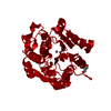
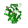
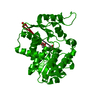
- PDB-4iu0: Crystal structure of Leishmania mexicana arginase in complex with... -
+ Open data

- Basic information
| Entry | Database: PDB / ID: 4iu0 | ||||||
|---|---|---|---|---|---|---|---|
| Title | Crystal structure of Leishmania mexicana arginase in complex with inhibitor ABH | ||||||
 Components Components | Arginase | ||||||
 Keywords Keywords | HYDROLASE/HYDROLASE INHIBITOR / arginase fold / HYDROLASE-HYDROLASE INHIBITOR complex | ||||||
| Function / homology |  Function and homology information Function and homology informationarginine metabolic process / arginase / arginase activity / urea cycle / manganese ion binding / nucleus / cytosol Similarity search - Function | ||||||
| Biological species |   Leishmania mexicana (eukaryote) Leishmania mexicana (eukaryote) | ||||||
| Method |  X-RAY DIFFRACTION / SYNCHROTRON / MOLECULAR REPLACEMENT / Resolution: 1.77 Å X-RAY DIFFRACTION / SYNCHROTRON / MOLECULAR REPLACEMENT / Resolution: 1.77 Å | ||||||
 Authors Authors | D'Antonio, E.L. / Ullman, B. / Roberts, S.C. / Gaur Dixit, U. / Wilson, M.E. / Hai, Y. / Christianson, D.W. | ||||||
 Citation Citation | Journal: Arch.Biochem.Biophys. / Year: 2013 Title: Crystal structure of arginase from Leishmania mexicana and implications for the inhibition of polyamine biosynthesis in parasitic infections. Authors: D'Antonio, E.L. / Ullman, B. / Roberts, S.C. / Dixit, U.G. / Wilson, M.E. / Hai, Y. / Christianson, D.W. | ||||||
| History |
|
- Structure visualization
| Structure viewer | Molecule: MolmilJmol/JSmol |
|---|
- Downloads & links
-Download
| PDBx/mmCIF format | 4iu0.cif.gz | 77.3 KB | Display | PDBx/mmCIF format |
|---|---|---|---|---|
| PDB format | pdb4iu0.ent.gz | 55.4 KB | Display | PDB format |
| PDBx/mmJSON format | 4iu0.json.gz | Tree view | PDBx/mmJSON format | |
| Others |  Other downloads Other downloads |
-Validation report
| Arichive directory | https://data.pdbj.org/pub/pdb/validation_reports/iu/4iu0ftp://data.pdbj.org/pub/pdb/validation_reports/iu/4iu0 | HTTPS FTP |
|---|
-Related structure data
| Related structure data |  4ityC  4iu1C 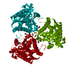 4iu4C 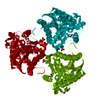 4iu5C 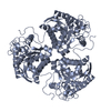 3tf3S C: citing same article ( S: Starting model for refinement |
|---|---|
| Similar structure data |


-Links
 PDBj
PDBj- Assembly
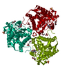
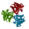
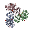
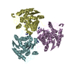
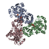
| Deposited unit | 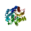
| ||||||||
|---|---|---|---|---|---|---|---|---|---|
| 1 | 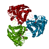
| ||||||||
| Unit cell |
|
-Components
| #1: Protein | Mass: 36034.449 Da / Num. of mol.: 1 / Fragment: UNP residues 13-329 Source method: isolated from a genetically manipulated source Source: (gene. exp.) Leishmania mexicana (eukaryote) / Gene: ARG / Plasmid: pET200/D-TOPO / Production host:  | ||||||
|---|---|---|---|---|---|---|---|
| #2: Chemical |   Mass: 54.938 Da / Num. of mol.: 2 / Source method: obtained synthetically / Formula: Mn Mass: 54.938 Da / Num. of mol.: 2 / Source method: obtained synthetically / Formula: Mn#3: Chemical | ChemComp-ABH / | 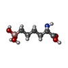  Mass: 191.998 Da / Num. of mol.: 1 / Source method: obtained synthetically / Formula: C6H15BNO5 Mass: 191.998 Da / Num. of mol.: 1 / Source method: obtained synthetically / Formula: C6H15BNO5#4: Chemical | ChemComp-GOL / |   Mass: 92.094 Da / Num. of mol.: 1 / Source method: obtained synthetically / Formula: C3H8O3 Mass: 92.094 Da / Num. of mol.: 1 / Source method: obtained synthetically / Formula: C3H8O3#5: Water | ChemComp-HOH / |  Mass: 18.015 Da / Num. of mol.: 81 / Source method: isolated from a natural source / Formula: H2O Mass: 18.015 Da / Num. of mol.: 81 / Source method: isolated from a natural source / Formula: H2O |
-Experimental details
-Experiment
| Experiment | Method: X-RAY DIFFRACTION / Number of used crystals: 1 |
|---|
- Sample preparation
| Crystal | Density Matthews: 2.44 Å3/Da / Density % sol: 49.63 % |
|---|---|
| Crystal grow | Temperature: 298 K / Method: vapor diffusion, hanging drop / pH: 7.5 Details: 5 uL protein solution (7.0 mg/mL delta-12-LmARG, 45 mM bicine, pH 8.5, 10 mM ABH, 90 uM manganese chloride, 4.5% v/v glycerol, 1.8 mM BME) + 5 uL precipitant solution (12% w/v PEG3350, 0.1 M ...Details: 5 uL protein solution (7.0 mg/mL delta-12-LmARG, 45 mM bicine, pH 8.5, 10 mM ABH, 90 uM manganese chloride, 4.5% v/v glycerol, 1.8 mM BME) + 5 uL precipitant solution (12% w/v PEG3350, 0.1 M HEPES, pH 7.5) equilibrated against 300 uL precipitant solution on a siliconized cover slide, VAPOR DIFFUSION, HANGING DROP, temperature 298K |
-Data collection
| Diffraction | Mean temperature: 100 K |
|---|---|
| Diffraction source | Source: SYNCHROTRON / Site: NSLS  / Beamline: X29A / Wavelength: 1.075 Å / Beamline: X29A / Wavelength: 1.075 Å |
| Detector | Type: ADSC QUANTUM 315 / Detector: CCD / Date: Jan 31, 2012 |
| Radiation | Monochromator: Cryogenically cooled double-crystal Si(111) with horizontally focusing, sagitally bent second crystal with 4:1 magnification ratio and vertically focusing mirror Protocol: SINGLE WAVELENGTH / Monochromatic (M) / Laue (L): M / Scattering type: x-ray |
| Radiation wavelength | Wavelength: 1.075 Å / Relative weight: 1 |
| Reflection twin | Operator: h,-h-k,-l / Fraction: 0.38 |
| Reflection | Resolution: 1.77→50 Å / Num. all: 33249 / Num. obs: 33141 / % possible obs: 100 % / Observed criterion σ(F): 0 / Observed criterion σ(I): -3 / Redundancy: 4.7 % / Rmerge(I) obs: 0.067 / Rsym value: 0.067 / Net I/σ(I): 21.896 |
| Reflection shell | Resolution: 1.77→1.83 Å / Redundancy: 4.7 % / Rmerge(I) obs: 0.699 / Mean I/σ(I) obs: 2.369 / Rsym value: 0.699 / % possible all: 100 |
- Processing
| Software |
| ||||||||||||||||||||||||||||||||||||
|---|---|---|---|---|---|---|---|---|---|---|---|---|---|---|---|---|---|---|---|---|---|---|---|---|---|---|---|---|---|---|---|---|---|---|---|---|---|
| Refinement | Method to determine structure: MOLECULAR REPLACEMENT Starting model: PDB ENTRY 3TF3 Resolution: 1.77→50 Å / Isotropic thermal model: ISOTROPIC / Cross valid method: THROUGHOUT / Stereochemistry target values: Engh & Huber
| ||||||||||||||||||||||||||||||||||||
| Solvent computation | Bsol: 56.9306 Å2 | ||||||||||||||||||||||||||||||||||||
| Displacement parameters | Biso mean: 29 Å2
| ||||||||||||||||||||||||||||||||||||
| Refine analyze |
| ||||||||||||||||||||||||||||||||||||
| Refinement step | Cycle: LAST / Resolution: 1.77→50 Å
| ||||||||||||||||||||||||||||||||||||
| Refine LS restraints |
| ||||||||||||||||||||||||||||||||||||
| LS refinement shell | Resolution: 1.77→1.83 Å /
| ||||||||||||||||||||||||||||||||||||
| Xplor file |
|
