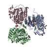
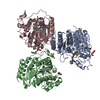
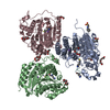
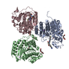
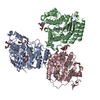
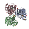
HYDROLASE / mixed alpha and beta structure / allophanate binding
Function / homology
Function and homology information
urea carboxylase / urea carboxylase activity / allophanate hydrolase / allophanate hydrolase activity / small molecule metabolic process / ATP binding / metal ion binding Similarity search - Function
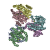
Methane Monooxygenase Hydroxylase; Chain G, domain 1 - #1700 / Urea carboxylase / Allophanate hydrolase / : / Allophanate hydrolase C-terminal domain / Carboxyltransferase domain, subdomain A and B / Carboxyltransferase domain, subdomain A and B / Allophanate hydrolase subunit 2 / Carboxyltransferase domain, subdomain C and D / Carboxyltransferase domain, subdomain C and D ...Methane Monooxygenase Hydroxylase; Chain G, domain 1 - #1700 / Urea carboxylase / Allophanate hydrolase / : / Allophanate hydrolase C-terminal domain / Carboxyltransferase domain, subdomain A and B / Carboxyltransferase domain, subdomain A and B / Allophanate hydrolase subunit 2 / Carboxyltransferase domain, subdomain C and D / Carboxyltransferase domain, subdomain C and D / Allophanate hydrolase subunit 1 / Gamma-glutamyl cyclotransferase-like / Hypothetical upf0131 protein ytfp / Amidase signature (AS) enzymes / Amidase signature (AS) domain / Amidase signature domain / Amidase signature (AS) superfamily / Amidase / : / Biotin-binding site / Biotin-requiring enzymes attachment site. / Biotin carboxylase-like, N-terminal domain / Biotin carboxylase, C-terminal / Biotin carboxylation domain / Biotin carboxylase, N-terminal domain / Biotin carboxylase C-terminal domain / Biotin carboxylation domain profile. / Biotin carboxylase C-terminal domain / Carbamoyl-phosphate synthase subdomain signature 1. / Carbamoyl-phosphate synthetase large subunit-like, ATP-binding domain / Carbamoyl-phosphate synthase L chain, ATP binding domain / Biotin-requiring enzyme / Rudiment single hybrid motif / Biotinyl/lipoyl domain profile. / Biotin/lipoyl attachment / Single hybrid motif / Pre-ATP-grasp domain superfamily / Cyclophilin-like domain superfamily / ATP-grasp fold / ATP-grasp fold profile. / Methane Monooxygenase Hydroxylase; Chain G, domain 1 / Carbamoyl-phosphate synthase subdomain signature 2. / Roll / Alpha-Beta Complex / Up-down Bundle / Mainly Alpha / Alpha Beta Similarity search - Domain/homology
Monochromator: double crystal Si(111) / Protocol: SINGLE WAVELENGTH / Monochromatic (M) / Laue (L): M / Scattering type: x-ray
Radiation wavelength
Wavelength: 0.979 Å / Relative weight: 1
Reflection
Resolution: 2.5→50 Å / Num. all: 53115 / Num. obs: 53061 / % possible obs: 99.9 % / Redundancy: 6 % / Rmerge(I) obs: 0.093 / Net I/σ(I): 6.1
Reflection shell
Resolution: 2.5→2.64 Å / Redundancy: 6 % / Rmerge(I) obs: 0.432 / Mean I/σ(I) obs: 1.6 / % possible all: 100
-
Processing
Software
Name
Version
Classification
HKL-2000
datacollection
SnB
phasing
REFMAC
5.6.0117
refinement
HKL-2000
datareduction
HKL-2000
datascaling
Refinement
Method to determine structure: SAD / Resolution: 2.5→46.53 Å / Cor.coef. Fo:Fc: 0.953 / Cor.coef. Fo:Fc free: 0.906 / SU B: 9.052 / SU ML: 0.197 / Cross valid method: THROUGHOUT / ESU R: 0.418 / ESU R Free: 0.277 / Stereochemistry target values: MAXIMUM LIKELIHOOD Details: HYDROGENS HAVE BEEN USED IF PRESENT IN THE INPUT U VALUES : REFINED INDIVIDUALLY
Rfactor
Num. reflection
% reflection
Selection details
Rfree
0.24659
2715
5.1 %
RANDOM
Rwork
0.17623
-
-
-
obs
0.17978
50331
99.85 %
-
all
-
50406
-
-
Solvent computation
Ion probe radii: 0.8 Å / Shrinkage radii: 0.8 Å / VDW probe radii: 1.2 Å / Solvent model: MASK
Displacement parameters
Biso mean: 38.517 Å2
Baniso -1
Baniso -2
Baniso -3
1-
0.28 Å2
0 Å2
0 Å2
2-
-
0.11 Å2
0 Å2
3-
-
-
-0.39 Å2
Refinement step
Cycle: LAST / Resolution: 2.5→46.53 Å
Protein
Nucleic acid
Ligand
Solvent
Total
Num. atoms
9479
0
16
273
9768
Refine LS restraints
Refine-ID
Type
Dev ideal
Dev ideal target
Number
X-RAY DIFFRACTION
r_bond_refined_d
0.015
0.02
9721
X-RAY DIFFRACTION
r_angle_refined_deg
1.977
1.983
13231
X-RAY DIFFRACTION
r_dihedral_angle_1_deg
7.759
5
1224
X-RAY DIFFRACTION
r_dihedral_angle_2_deg
39.341
24.785
395
X-RAY DIFFRACTION
r_dihedral_angle_3_deg
21.213
15
1600
X-RAY DIFFRACTION
r_dihedral_angle_4_deg
22.093
15
40
X-RAY DIFFRACTION
r_chiral_restr
0.147
0.2
1497
X-RAY DIFFRACTION
r_gen_planes_refined
0.008
0.021
7332
Refine LS restraints NCS
Dom-ID: 1 / Auth asym-ID: A / Refine-ID: X-RAY DIFFRACTION
Ens-ID
Number
Type
Rms dev position (Å)
Weight position
1
3691
MEDIUMPOSITIONAL
0.35
0.5
1
3691
MEDIUMTHERMAL
4.81
2
2
1040
MEDIUMPOSITIONAL
0.47
0.5
2
1040
MEDIUMTHERMAL
8.8
2
LS refinement shell
Resolution: 2.5→2.565 Å / Total num. of bins used: 20
Rfactor
Num. reflection
% reflection
Rfree
0.363
196
-
Rwork
0.262
3387
-
obs
-
-
99.97 %
+
About Yorodumi
-
News
-
Feb 9, 2022. New format data for meta-information of EMDB entries
New format data for meta-information of EMDB entries
Version 3 of the EMDB header file is now the official format.
The previous official version 1.9 will be removed from the archive.
In the structure databanks used in Yorodumi, some data are registered as the other names, "COVID-19 virus" and "2019-nCoV". Here are the details of the virus and the list of structure data.
Jan 31, 2019. EMDB accession codes are about to change! (news from PDBe EMDB page)
EMDB accession codes are about to change! (news from PDBe EMDB page)
The allocation of 4 digits for EMDB accession codes will soon come to an end. Whilst these codes will remain in use, new EMDB accession codes will include an additional digit and will expand incrementally as the available range of codes is exhausted. The current 4-digit format prefixed with “EMD-” (i.e. EMD-XXXX) will advance to a 5-digit format (i.e. EMD-XXXXX), and so on. It is currently estimated that the 4-digit codes will be depleted around Spring 2019, at which point the 5-digit format will come into force.
The EM Navigator/Yorodumi systems omit the EMD- prefix.
Related info.:Q: What is EMD? / ID/Accession-code notation in Yorodumi/EM Navigator
Yorodumi is a browser for structure data from EMDB, PDB, SASBDB, etc.
This page is also the successor to EM Navigator detail page, and also detail information page/front-end page for Omokage search.
The word "yorodu" (or yorozu) is an old Japanese word meaning "ten thousand". "mi" (miru) is to see.
Related info.:EMDB / PDB / SASBDB / Comparison of 3 databanks / Yorodumi Search / Aug 31, 2016. New EM Navigator & Yorodumi / Yorodumi Papers / Jmol/JSmol / Function and homology information / Changes in new EM Navigator and Yorodumi
 Movie
Movie Controller


 Open data
 Basic information
 Components
 Keywords
 Function and homology information
 Kluyveromyces lactis (yeast)
 X-RAY DIFFRACTION /
  Authors
 Citation
 Structure visualization
 Downloads & links
 Other downloads
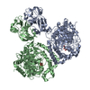


 PDBj
PDBj


 Assembly
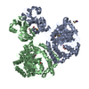


 Mass: 150.087 Da / Num. of mol.: 1 / Source method: obtained synthetically / Formula: C4H6O6
 Mass: 92.094 Da / Num. of mol.: 1 / Source method: obtained synthetically / Formula: C3H8O3
 Mass: 18.015 Da / Num. of mol.: 273 / Source method: isolated from a natural source / Formula: H2O
 Sample preparation
 / Beamline: BL17U / Wavelength: 0.979 Å
 Processing