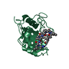
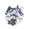
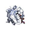
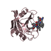
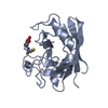
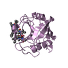
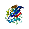
Entry Database : PDB / ID : 4ipzTitle SmBz bound to Cyclophilin A Peptidyl-prolyl cis-trans isomerase A cyclosporine SmBz-CsA Keywords / / / / Function / homology Function Domain/homology Component
/ / / / / / / / / / / / / / / / / / / / / / / / / / / / / / / / / / / / / / / / / / / / / / / / / / / / / / / / / / / / / / / / / / / / / / / / / / / / / / / / Biological species Homo sapiens (human)Tolypocladium inflatum (fungus)Method / / / Resolution : 1.67 Å Authors Price, A.J. / Jacques, D.A. / James, L.C. Journal : Nature / Year : 2013Title : HIV-1 evades innate immune recognition through specific cofactor recruitment.Authors : Rasaiyaah, J. / Tan, C.P. / Fletcher, A.J. / Price, A.J. / Blondeau, C. / Hilditch, L. / Jacques, D.A. / Selwood, D.L. / James, L.C. / Noursadeghi, M. / Towers, G.J. History Deposition Jan 10, 2013 Deposition site / Processing site Revision 1.0 Nov 6, 2013 Provider / Type Revision 1.1 Dec 4, 2013 Group Revision 1.2 Aug 29, 2018 Group / Source and taxonomy / Structure summaryCategory / entity_src_nat / pdbx_entity_src_synItem / _entity.pdbx_mutation / _entity.src_methodRevision 1.3 Sep 20, 2023 Group Data collection / Database references ... Data collection / Database references / Derived calculations / Refinement description Category chem_comp_atom / chem_comp_bond ... chem_comp_atom / chem_comp_bond / database_2 / pdbx_initial_refinement_model / struct_conn / struct_ref_seq_dif / struct_site Item _database_2.pdbx_DOI / _database_2.pdbx_database_accession ... _database_2.pdbx_DOI / _database_2.pdbx_database_accession / _struct_conn.pdbx_dist_value / _struct_conn.pdbx_leaving_atom_flag / _struct_conn.ptnr1_auth_comp_id / _struct_conn.ptnr1_auth_seq_id / _struct_conn.ptnr1_label_atom_id / _struct_conn.ptnr1_label_comp_id / _struct_conn.ptnr1_label_seq_id / _struct_conn.ptnr2_auth_comp_id / _struct_conn.ptnr2_auth_seq_id / _struct_conn.ptnr2_label_atom_id / _struct_conn.ptnr2_label_comp_id / _struct_conn.ptnr2_label_seq_id / _struct_ref_seq_dif.details / _struct_site.pdbx_auth_asym_id / _struct_site.pdbx_auth_comp_id / _struct_site.pdbx_auth_seq_id Revision 2.0 Nov 15, 2023 Group / Data collection / Derived calculationsCategory atom_site / atom_site_anisotrop ... atom_site / atom_site_anisotrop / chem_comp_atom / chem_comp_bond / struct_conn Item _atom_site.auth_atom_id / _atom_site.label_atom_id ... _atom_site.auth_atom_id / _atom_site.label_atom_id / _atom_site_anisotrop.pdbx_auth_atom_id / _atom_site_anisotrop.pdbx_label_atom_id / _chem_comp_atom.atom_id / _chem_comp_bond.atom_id_1 / _chem_comp_bond.atom_id_2 / _struct_conn.pdbx_leaving_atom_flag Revision 3.0 May 8, 2024 Group / Category / atom_site_anisotropItem _atom_site.B_iso_or_equiv / _atom_site.Cartn_x ... _atom_site.B_iso_or_equiv / _atom_site.Cartn_x / _atom_site.Cartn_y / _atom_site.Cartn_z / _atom_site.auth_atom_id / _atom_site.label_atom_id / _atom_site.type_symbol / _atom_site_anisotrop.U[1][1] / _atom_site_anisotrop.U[1][2] / _atom_site_anisotrop.U[1][3] / _atom_site_anisotrop.U[2][2] / _atom_site_anisotrop.U[2][3] / _atom_site_anisotrop.U[3][3] / _atom_site_anisotrop.pdbx_auth_atom_id / _atom_site_anisotrop.pdbx_label_atom_id / _atom_site_anisotrop.type_symbol
Show all Show less
 Movie
Movie Controller


 Open data
 Basic information
 Components
 Keywords
 Function and homology information
 Homo sapiens (human)
 Tolypocladium inflatum (fungus)
 X-RAY DIFFRACTION /
  Authors
 Citation
 Structure visualization
 Downloads & links
 Other downloads
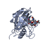

 PDBj
PDBj


 Assembly


 Type: Cyclic peptide / Class: Immunosuppressant / Mass: 1354.757 Da / Num. of mol.: 1 / Mutation: (sar)7(1JM) / Source method: obtained synthetically / Details: CsA has been modified at position 4 (sarcosine) / Source: (synth.)
 Mass: 35.453 Da / Num. of mol.: 2 / Source method: obtained synthetically / Formula: Cl
 Mass: 18.015 Da / Num. of mol.: 184 / Source method: isolated from a natural source / Formula: H2O
 Sample preparation
 Processing