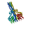
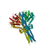
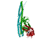
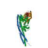
[English] 日本語
Yorodumi
- PDB-4im0: Structure of Tank-Binding Kinase 1 -

+
Open data


ID or keywords:

Loading...

-
Basic information

Entry
Database: PDB / ID: 4im0
TitleStructure of Tank-Binding Kinase 1
ComponentsSerine/threonine-protein kinase TBK1
KeywordsTRANSFERASE/TRANSFERASE INHIBITOR / kinase / serine/threonine kinase / MRT67307 / TRANSFERASE-TRANSFERASE INHIBITOR complex
Function / homology
Function and homology information


IRF3 mediated activation of type 1 IFN / STAT6-mediated induction of chemokines / positive regulation of xenophagy / serine/threonine protein kinase complex / regulation of type I interferon production / dendritic cell proliferation / IRF3-mediated induction of type I IFN / positive regulation of type I interferon-mediated signaling pathway / Interleukin-37 signaling / TNFR1-induced proapoptotic signaling ...IRF3 mediated activation of type 1 IFN / STAT6-mediated induction of chemokines / positive regulation of xenophagy / serine/threonine protein kinase complex / regulation of type I interferon production / dendritic cell proliferation / IRF3-mediated induction of type I IFN / positive regulation of type I interferon-mediated signaling pathway / Interleukin-37 signaling / TNFR1-induced proapoptotic signaling / TRAF6 mediated IRF7 activation / type I interferon-mediated signaling pathway / cytoplasmic pattern recognition receptor signaling pathway / positive regulation of interferon-alpha production / antiviral innate immune response / positive regulation of macroautophagy / positive regulation of type I interferon production / canonical NF-kappaB signal transduction / positive regulation of autophagy / negative regulation of TORC1 signaling / TICAM1-dependent activation of IRF3/IRF7 / positive regulation of TORC1 signaling / Regulation of innate immune responses to cytosolic DNA / positive regulation of interferon-beta production / activation of innate immune response / Activation of IRF3, IRF7 mediated by TBK1, IKKε (IKBKE) / Negative regulators of DDX58/IFIH1 signaling / Regulation of TNFR1 signaling / phosphoprotein binding / peptidyl-threonine phosphorylation / response to virus / DDX58/IFIH1-mediated induction of interferon-alpha/beta / SARS-CoV-1 activates/modulates innate immune responses / positive regulation of peptidyl-serine phosphorylation / TRAF3-dependent IRF activation pathway / defense response to virus / protein phosphatase binding / peptidyl-serine phosphorylation / positive regulation of canonical NF-kappaB signal transduction / Potential therapeutics for SARS / nucleic acid binding / non-specific serine/threonine protein kinase / protein kinase activity / defense response to Gram-positive bacterium / inflammatory response / negative regulation of gene expression / protein phosphorylation / protein serine kinase activity / innate immune response / intracellular membrane-bounded organelle / protein serine/threonine kinase activity / SARS-CoV-2 activates/modulates innate and adaptive immune responses / positive regulation of transcription by RNA polymerase II / nucleoplasm / ATP binding / identical protein binding / cytosol / cytoplasm
Similarity search - Function
Substrate Binding Domain Of Dnak; Chain:A; Domain 2 - #420 / TANK binding kinase 1, ubiquitin-like domain / TANK-binding kinase 1, coiled-coil domain 1 / TANK-binding kinase 1 coiled-coil domain 1 / TANK binding kinase 1 ubiquitin-like domain / Substrate Binding Domain Of Dnak; Chain:A; Domain 2 / Phosphatidylinositol 3-kinase Catalytic Subunit; Chain A, domain 1 / Ubiquitin-like (UB roll) / Transferase(Phosphotransferase) domain 1 / Transferase(Phosphotransferase); domain 1 ...Substrate Binding Domain Of Dnak; Chain:A; Domain 2 - #420 / TANK binding kinase 1, ubiquitin-like domain / TANK-binding kinase 1, coiled-coil domain 1 / TANK-binding kinase 1 coiled-coil domain 1 / TANK binding kinase 1 ubiquitin-like domain / Substrate Binding Domain Of Dnak; Chain:A; Domain 2 / Phosphatidylinositol 3-kinase Catalytic Subunit; Chain A, domain 1 / Ubiquitin-like (UB roll) / Transferase(Phosphotransferase) domain 1 / Transferase(Phosphotransferase); domain 1 / Phosphorylase Kinase; domain 1 / Phosphorylase Kinase; domain 1 / Protein kinase domain / Serine/Threonine protein kinases, catalytic domain / Roll / Protein kinase, ATP binding site / Protein kinases ATP-binding region signature. / Protein kinase domain profile. / Protein kinase domain / Protein kinase-like domain superfamily / Up-down Bundle / 2-Layer Sandwich / Orthogonal Bundle / Mainly Alpha / Alpha Beta
Similarity search - Domain/homology
Chem-1FV / Serine/threonine-protein kinase TBK1
Similarity search - Component
Biological speciesHomo sapiens (human)
MethodX-RAY DIFFRACTION / SYNCHROTRON / SAD / Resolution: 2.4001 Å
AuthorsTu, D. / Eck, M.J.
CitationJournal: Cell Rep / Year: 2013
Title: Structure and ubiquitination-dependent activation of TANK-binding kinase 1.
Authors: Tu, D. / Zhu, Z. / Zhou, A.Y. / Yun, C.H. / Lee, K.E. / Toms, A.V. / Li, Y. / Dunn, G.P. / Chan, E. / Thai, T. / Yang, S. / Ficarro, S.B. / Marto, J.A. / Jeon, H. / Hahn, W.C. / Barbie, D.A. / Eck, M.J.
History
DepositionJan 1, 2013Deposition site: RCSB / Processing site: RCSB
Revision 1.0Mar 6, 2013Provider: repository / Type: Initial release
Revision 1.1Mar 20, 2013Group: Other
Revision 1.2Jul 17, 2019Group: Data collection / Database references / Category: citation / citation_author
Item: _citation.country / _citation.journal_abbrev ..._citation.country / _citation.journal_abbrev / _citation.journal_id_CSD / _citation.journal_id_ISSN / _citation.journal_volume / _citation.page_first / _citation.page_last / _citation.pdbx_database_id_DOI / _citation.pdbx_database_id_PubMed / _citation.title / _citation_author.name
Revision 1.3Feb 28, 2024Group: Data collection / Database references / Derived calculations
Category: chem_comp_atom / chem_comp_bond ...chem_comp_atom / chem_comp_bond / database_2 / struct_ref_seq_dif / struct_site
Item: _database_2.pdbx_DOI / _database_2.pdbx_database_accession ..._database_2.pdbx_DOI / _database_2.pdbx_database_accession / _struct_ref_seq_dif.details / _struct_site.pdbx_auth_asym_id / _struct_site.pdbx_auth_comp_id / _struct_site.pdbx_auth_seq_id

-
Structure visualization

Structure viewerMolecule:
MolmilJmol/JSmol

Downloads & links

-
Assembly

Deposited unit
A: Serine/threonine-protein kinase TBK1
hetero molecules


Theoretical massNumber of molelcules
Total (without water)76,6502
Polymers76,1851
Non-polymers4651
Water5,260292
1
A: Serine/threonine-protein kinase TBK1
hetero molecules

A: Serine/threonine-protein kinase TBK1
hetero molecules


Theoretical massNumber of molelcules
Total (without water)153,2994
Polymers152,3702
Non-polymers9292
Water362
TypeNameSymmetry operationNumber
identity operation1_555x,y,z1
crystal symmetry operation6_764-x+2,-x+y+1,-z-1/31
Buried area3080 Å2
ΔGint-2 kcal/mol
Surface area61560 Å2
MethodPISA
Unit cell
Length a, b, c (Å)133.921, 133.921, 85.670
Angle α, β, γ (deg.)90.00, 90.00, 120.00
Int Tables number154
Space group name H-MP3221
Components on special symmetry positions
IDModelComponents
11A-852-

HOH

-
Components

#1: Protein Serine/threonine-protein kinase TBK1 / NF-kappa-B-activating kinase / T2K / TANK-binding kinase 1


Mass: 76185.062 Da / Num. of mol.: 1 / Fragment: residues 1 to 657 / Mutation: D135N
Source method: isolated from a genetically manipulated source
Source: (gene. exp.) Homo sapiens (human) / Gene: TBK1, NAK / Production host: Spodoptera frugiperda (fall armyworm)
References: UniProt: Q9UHD2, non-specific serine/threonine protein kinase
#2: Chemical ChemComp-1FV / N-{3-[(5-cyclopropyl-2-{[3-(morpholin-4-ylmethyl)phenyl]amino}pyrimidin-4-yl)amino]propyl}cyclobutanecarboxamide / MRT67307


Mass: 464.603 Da / Num. of mol.: 1 / Source method: obtained synthetically / Formula: C26H36N6O2
#3: Water ChemComp-HOH / water / Water


Mass: 18.015 Da / Num. of mol.: 292 / Source method: isolated from a natural source / Formula: H2O

-
Experimental details

-
Experiment

ExperimentMethod: X-RAY DIFFRACTION / Number of used crystals: 2

-
Sample preparation

CrystalDensity Matthews: 2.91 Å3/Da / Density % sol: 57.75 %
Crystal growTemperature: 293 K / Method: vapor diffusion, hanging drop / pH: 8.7
Details: 4% (w/v) PEG8000, 100 mM Tris pH 8.7, VAPOR DIFFUSION, HANGING DROP, temperature 293K

-
Data collection

DiffractionMean temperature: 100 K
Diffraction sourceSource: SYNCHROTRON / Site: APS / Beamline: 24-ID-E / Wavelength: 0.97918 Å
DetectorType: ADSC QUANTUM 315 / Detector: CCD / Date: Jun 30, 2011
RadiationMonochromator: MD2 micro-diffractometer / Protocol: SINGLE WAVELENGTH / Monochromatic (M) / Laue (L): M / Scattering type: x-ray
Radiation wavelengthWavelength: 0.97918 Å / Relative weight: 1
ReflectionResolution: 2.4→50 Å / Num. all: 35090 / Num. obs: 34873 / % possible obs: 99.4 % / Observed criterion σ(F): 0 / Observed criterion σ(I): -3
Reflection shell
Resolution (Å)Diffraction-ID% possible all
2.4-2.55199.6
2.55-2.75199.6
2.75-3.02199.3
3.02-3.461100
3.46-4.361100
4.36-50197.8

-
Processing

Software
NameVersionClassification
HKL-2000data collection
SHARPphasing
PHENIX(phenix.refine: 1.7.3_928)refinement
HKL-2000data reduction
HKL-2000data scaling
RefinementMethod to determine structure: SAD / Resolution: 2.4001→39.024 Å / SU ML: 0.38 / σ(F): 0 / Phase error: 27.22 / Stereochemistry target values: ML
RfactorNum. reflection% reflectionSelection details
Rfree0.2357 1748 5.04 %random
Rwork0.1993 ---
all0.2012 34940 --
obs0.2012 34682 99.26 %-
Solvent computationShrinkage radii: 0.98 Å / VDW probe radii: 1.2 Å / Solvent model: FLAT BULK SOLVENT MODEL / Bsol: 74.922 Å2 / ksol: 0.304 e/Å3
Displacement parameters
Baniso -1Baniso -2Baniso -3
1--4.0634 Å20 Å20 Å2
2---4.0634 Å20 Å2
3---8.1267 Å2
Refinement stepCycle: LAST / Resolution: 2.4001→39.024 Å
ProteinNucleic acidLigandSolventTotal
Num. atoms4931 0 34 292 5257
Refine LS restraints
Refine-IDTypeDev idealNumber
X-RAY DIFFRACTIONf_bond_d0.0055088
X-RAY DIFFRACTIONf_angle_d0.8896879
X-RAY DIFFRACTIONf_dihedral_angle_d15.6211895
X-RAY DIFFRACTIONf_chiral_restr0.055768
X-RAY DIFFRACTIONf_plane_restr0.003877
LS refinement shell
Resolution (Å)Rfactor RfreeNum. reflection RfreeRfactor RworkNum. reflection RworkRefine-ID% reflection obs (%)
2.4001-2.47070.34141220.27212733X-RAY DIFFRACTION100
2.4707-2.55040.27051500.25852742X-RAY DIFFRACTION100
2.5504-2.64160.32291480.26012702X-RAY DIFFRACTION100
2.6416-2.74730.36751390.26242757X-RAY DIFFRACTION100
2.7473-2.87230.35831470.26042696X-RAY DIFFRACTION99
2.8723-3.02370.35261500.24632724X-RAY DIFFRACTION99
3.0237-3.21310.30381550.2412716X-RAY DIFFRACTION100
3.2131-3.4610.25671470.22292766X-RAY DIFFRACTION100
3.461-3.8090.2081530.19472756X-RAY DIFFRACTION100
3.809-4.35960.20471420.16922768X-RAY DIFFRACTION100
4.3596-5.49020.22011520.16862788X-RAY DIFFRACTION99
5.4902-39.02910.18981430.18682786X-RAY DIFFRACTION96
Refinement TLS params.

Method: refined / Refine-ID: X-RAY DIFFRACTION

IDL112)L122)L132)L222)L232)L332)S11 (Å °)S12 (Å °)S13 (Å °)S21 (Å °)S22 (Å °)S23 (Å °)S31 (Å °)S32 (Å °)S33 (Å °)T112)T122)T132)T222)T232)T332)Origin x (Å)Origin y (Å)Origin z (Å)
14.3907-1.0355-1.10473.12491.692.56440.03370.3216-1.3509-0.5632-0.12870.10940.6389-0.01390.04940.80150.06040.04770.402-0.01050.7962103.551216.0611-31.7997
26.44822.7475-0.68485.871-3.5569.0619-0.3051-0.7618-1.4526-0.16640.25860.91341.5749-0.4627-0.00310.64630.00130.1640.5830.14331.002584.028515.8643-12.2129
32.7522-0.0020.04633.88871.00251.5366-0.0391-0.06350.0371-0.2459-0.0160.6124-0.2242-0.37560.03160.36950.06690.02950.43310.06150.445588.806745.022-14.6174
48.9259-7.2085-0.4715.37090.3175-0.0747-1.1766-0.41650.67521.84720.6321-0.1345-0.645-0.70360.67271.2250.5266-0.29461.1408-0.33810.969273.347289.1813-1.8101
54.6946-0.61020.88814.4069-0.58842.2223-0.1348-0.5003-0.62670.38650.2621-0.8870.44810.11340.05650.37310.0860.09590.40210.27520.2964108.163129.52430.6272
63.7141-4.44093.44074.1857-3.19521.6077-0.7404-1.09070.52410.33520.59160.389-0.4506-0.27080.00440.80170.30350.03930.7537-0.03690.894778.674570.2234-5.0842
Refinement TLS group
IDRefine-IDRefine TLS-IDSelection details
1X-RAY DIFFRACTION1chain 'A' and (resseq -1:216)
2X-RAY DIFFRACTION2chain 'A' and (resseq 217:270)
3X-RAY DIFFRACTION3chain 'A' and (resseq 271:470)
4X-RAY DIFFRACTION4chain 'A' and (resseq 471:526)
5X-RAY DIFFRACTION5chain 'A' and (resseq 527:604)
6X-RAY DIFFRACTION6chain 'A' and (resseq 605:657)

+
About Yorodumi

-
News

-
Feb 9, 2022. New format data for meta-information of EMDB entries

New format data for meta-information of EMDB entries

  • Version 3 of the EMDB header file is now the official format.
  • The previous official version 1.9 will be removed from the archive.

Related info.:EMDB header

External links:wwPDB to switch to version 3 of the EMDB data model

-
Aug 12, 2020. Covid-19 info

Covid-19 info

URL: https://pdbj.org/emnavi/covid19.php

New page: Covid-19 featured information page in EM Navigator.

Related info.:Covid-19 info / Mar 5, 2020. Novel coronavirus structure data

+
Mar 5, 2020. Novel coronavirus structure data

Novel coronavirus structure data

Related info.:Yorodumi Speices / Aug 12, 2020. Covid-19 info

External links:COVID-19 featured content - PDBj / Molecule of the Month (242):Coronavirus Proteases

+
Jan 31, 2019. EMDB accession codes are about to change! (news from PDBe EMDB page)

EMDB accession codes are about to change! (news from PDBe EMDB page)

  • The allocation of 4 digits for EMDB accession codes will soon come to an end. Whilst these codes will remain in use, new EMDB accession codes will include an additional digit and will expand incrementally as the available range of codes is exhausted. The current 4-digit format prefixed with “EMD-” (i.e. EMD-XXXX) will advance to a 5-digit format (i.e. EMD-XXXXX), and so on. It is currently estimated that the 4-digit codes will be depleted around Spring 2019, at which point the 5-digit format will come into force.
  • The EM Navigator/Yorodumi systems omit the EMD- prefix.

Related info.:Q: What is EMD? / ID/Accession-code notation in Yorodumi/EM Navigator

External links:EMDB Accession Codes are Changing Soon! / Contact to PDBj

+
Jul 12, 2017. Major update of PDB

Major update of PDB

  • wwPDB released updated PDB data conforming to the new PDBx/mmCIF dictionary.
  • This is a major update changing the version number from 4 to 5, and with Remediation, in which all the entries are updated.
  • In this update, many items about electron microscopy experimental information are reorganized (e.g. em_software).
  • Now, EM Navigator and Yorodumi are based on the updated data.

External links:wwPDB Remediation / Enriched Model Files Conforming to OneDep Data Standards Now Available in the PDB FTP Archive

-
Yorodumi

Thousand views of thousand structures

  • Yorodumi is a browser for structure data from EMDB, PDB, SASBDB, etc.
  • This page is also the successor to EM Navigator detail page, and also detail information page/front-end page for Omokage search.
  • The word "yorodu" (or yorozu) is an old Japanese word meaning "ten thousand". "mi" (miru) is to see.

Related info.:EMDB / PDB / SASBDB / Comparison of 3 databanks / Yorodumi Search / Aug 31, 2016. New EM Navigator & Yorodumi / Yorodumi Papers / Jmol/JSmol / Function and homology information / Changes in new EM Navigator and Yorodumi

Read more