 Movie
Movie Controller

[English] 日本語
 Yorodumi
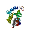
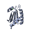
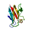
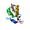
- PDB-4iej: Crystal structure of a DNA methyltransferase 1 associated protein... -
+ Open data

- Basic information
| Entry | Database: PDB / ID: 4iej | ||||||
|---|---|---|---|---|---|---|---|
| Title | Crystal structure of a DNA methyltransferase 1 associated protein 1 (DMAP1) from Homo sapiens at 1.45 A resolution | ||||||
 Components Components | DNA methyltransferase 1-associated protein 1 | ||||||
 Keywords Keywords | TRANSCRIPTION / DNA methylation / chromatin regulator / repressor / Structural Genomics / Joint Center for Structural Genomics / JCSG / Partnership for T-Cell Biology / TCELL / Protein Structure Initiative / PSI-BIOLOGY | ||||||
| Function / homology |  Function and homology information Function and homology informationSwr1 complex / regulation of double-strand break repair / NuA4 histone acetyltransferase complex / positive regulation of double-strand break repair via homologous recombination / replication fork / positive regulation of protein import into nucleus / transcription corepressor activity / nucleosome / HATs acetylate histones / regulation of apoptotic process ...Swr1 complex / regulation of double-strand break repair / NuA4 histone acetyltransferase complex / positive regulation of double-strand break repair via homologous recombination / replication fork / positive regulation of protein import into nucleus / transcription corepressor activity / nucleosome / HATs acetylate histones / regulation of apoptotic process / RNA polymerase II-specific DNA-binding transcription factor binding / response to ethanol / regulation of cell cycle / chromatin remodeling / negative regulation of DNA-templated transcription / DNA repair / regulation of DNA-templated transcription / positive regulation of DNA-templated transcription / negative regulation of transcription by RNA polymerase II / nucleoplasm / nucleus / cytosol / cytoplasm Similarity search - Function | ||||||
| Biological species |  Homo sapiens (human) Homo sapiens (human) | ||||||
| Method |  X-RAY DIFFRACTION / SYNCHROTRON / MOLECULAR REPLACEMENT / molecular replacement / Resolution: 1.45 Å X-RAY DIFFRACTION / SYNCHROTRON / MOLECULAR REPLACEMENT / molecular replacement / Resolution: 1.45 Å | ||||||
 Authors Authors | Joint Center for Structural Genomics (JCSG) / Partnership for T-Cell Biology (TCELL) | ||||||
 Citation Citation | Journal: To be published Title: Crystal structure of a DNA methyltransferase 1 associated protein 1 (DMAP1) from Homo sapiens at 1.45 A resolution Authors: Joint Center for Structural Genomics (JCSG) / Partnership for T-Cell Biology | ||||||
| History |
|
- Structure visualization
| Structure viewer | Molecule: MolmilJmol/JSmol |
|---|
- Downloads & links
-Download
| PDBx/mmCIF format | 4iej.cif.gz | 52.9 KB | Display | PDBx/mmCIF format |
|---|---|---|---|---|
| PDB format | pdb4iej.ent.gz | 36.7 KB | Display | PDB format |
| PDBx/mmJSON format | 4iej.json.gz | Tree view | PDBx/mmJSON format | |
| Others |  Other downloads Other downloads |
-Validation report
| Arichive directory | https://data.pdbj.org/pub/pdb/validation_reports/ie/4iejftp://data.pdbj.org/pub/pdb/validation_reports/ie/4iej | HTTPS FTP |
|---|
-Related structure data
| Related structure data |  3hm5S S: Starting model for refinement |
|---|---|
| Similar structure data | |
| Other databases |


-Links
 PDBj
PDBj- Assembly
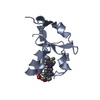
| Deposited unit | 
| ||||||||
|---|---|---|---|---|---|---|---|---|---|
| 1 |
| ||||||||
| Unit cell |
| ||||||||
| Components on special symmetry positions |
|
-Components
| #1: Protein | Mass: 11216.549 Da / Num. of mol.: 1 / Fragment: SANT domain containing residues 121-212 Source method: isolated from a genetically manipulated source Source: (gene. exp.) Homo sapiens (human) / Gene: BC008053, DMAP1, KIAA1425 / Plasmid: SpeedET / Production host:  |
|---|---|
| #2: Chemical | ChemComp-CA /   Mass: 40.078 Da / Num. of mol.: 1 / Source method: obtained synthetically / Formula: Ca Mass: 40.078 Da / Num. of mol.: 1 / Source method: obtained synthetically / Formula: Ca |
| #3: Water | ChemComp-HOH /  Mass: 18.015 Da / Num. of mol.: 76 / Source method: isolated from a natural source / Formula: H2O Mass: 18.015 Da / Num. of mol.: 76 / Source method: isolated from a natural source / Formula: H2O |
| Sequence details | THE CONSTRUCT (121-212) WAS EXPRESSED WITH A PURIFICATION TAG MGSDKIHHHHHHENLYFQG. THE TAG WAS ...THE CONSTRUCT (121-212) WAS EXPRESSED WITH A PURIFICATI |
-Experimental details
-Experiment
| Experiment | Method: X-RAY DIFFRACTION / Number of used crystals: 1 |
|---|
- Sample preparation
| Crystal | Density Matthews: 2.06 Å3/Da / Density % sol: 40.42 % |
|---|---|
| Crystal grow | Temperature: 277 K / Method: vapor diffusion, sitting drop Details: 0.20M calcium chloride 20.00% polyethylene glycol 3350, NANODROP, VAPOR DIFFUSION, SITTING DROP, temperature 277K |
-Data collection
| Diffraction | Mean temperature: 100 K | |||||||||||||||||||||||||||||||||||||||||||||||||||||||||||||||||||||||||||||
|---|---|---|---|---|---|---|---|---|---|---|---|---|---|---|---|---|---|---|---|---|---|---|---|---|---|---|---|---|---|---|---|---|---|---|---|---|---|---|---|---|---|---|---|---|---|---|---|---|---|---|---|---|---|---|---|---|---|---|---|---|---|---|---|---|---|---|---|---|---|---|---|---|---|---|---|---|---|---|
| Diffraction source | Source: SYNCHROTRON / Site: SSRL  / Beamline: BL12-2 / Wavelength: 0.9795 / Beamline: BL12-2 / Wavelength: 0.9795 | |||||||||||||||||||||||||||||||||||||||||||||||||||||||||||||||||||||||||||||
| Detector | Type: DECTRIS PILATUS 6M / Detector: PIXEL / Date: May 31, 2012 Details: Rhodium-coated vertical and horizontal focusing mirrors, liquid-nitrogen cooled double crystal Si(111) monochromator | |||||||||||||||||||||||||||||||||||||||||||||||||||||||||||||||||||||||||||||
| Radiation | Monochromator: double crystal Si(111) / Protocol: SINGLE WAVELENGTH / Monochromatic (M) / Laue (L): M / Scattering type: x-ray | |||||||||||||||||||||||||||||||||||||||||||||||||||||||||||||||||||||||||||||
| Radiation wavelength | Wavelength: 0.9795 Å / Relative weight: 1 | |||||||||||||||||||||||||||||||||||||||||||||||||||||||||||||||||||||||||||||
| Reflection | Resolution: 1.45→38.327 Å / Num. obs: 18199 / % possible obs: 99.7 % / Observed criterion σ(I): -3 / Biso Wilson estimate: 23.311 Å2 / Rmerge(I) obs: 0.05 / Net I/σ(I): 18.99 | |||||||||||||||||||||||||||||||||||||||||||||||||||||||||||||||||||||||||||||
| Reflection shell |
|
-Phasing
| Phasing | Method: molecular replacement |
|---|
- Processing
| Software |
| ||||||||||||||||||||||||||||||||||||||||||||||||||||||||||||||||||||||||||||||||||||||||||||||||||||||||||||
|---|---|---|---|---|---|---|---|---|---|---|---|---|---|---|---|---|---|---|---|---|---|---|---|---|---|---|---|---|---|---|---|---|---|---|---|---|---|---|---|---|---|---|---|---|---|---|---|---|---|---|---|---|---|---|---|---|---|---|---|---|---|---|---|---|---|---|---|---|---|---|---|---|---|---|---|---|---|---|---|---|---|---|---|---|---|---|---|---|---|---|---|---|---|---|---|---|---|---|---|---|---|---|---|---|---|---|---|---|---|
| Refinement | Method to determine structure: MOLECULAR REPLACEMENT Starting model: 3hm5 Resolution: 1.45→38.327 Å / Cor.coef. Fo:Fc: 0.9512 / Cor.coef. Fo:Fc free: 0.9482 / Occupancy max: 1 / Occupancy min: 0.3 / Cross valid method: THROUGHOUT / σ(F): 0 Details: 1. ATOM RECORD CONTAINS SUM OF TLS AND RESIDUAL B FACTORS. ANISOU RECORD CONTAINS SUM OF TLS AND RESIDUAL U FACTORS. 2. CALCIUM ION MODELED IS PRESENT IN CRYSTALLIZATION BUFFER.
| ||||||||||||||||||||||||||||||||||||||||||||||||||||||||||||||||||||||||||||||||||||||||||||||||||||||||||||
| Displacement parameters | Biso max: 112.38 Å2 / Biso mean: 36.2049 Å2 / Biso min: 17.68 Å2
| ||||||||||||||||||||||||||||||||||||||||||||||||||||||||||||||||||||||||||||||||||||||||||||||||||||||||||||
| Refine analyze | Luzzati coordinate error obs: 0.251 Å | ||||||||||||||||||||||||||||||||||||||||||||||||||||||||||||||||||||||||||||||||||||||||||||||||||||||||||||
| Refinement step | Cycle: LAST / Resolution: 1.45→38.327 Å
| ||||||||||||||||||||||||||||||||||||||||||||||||||||||||||||||||||||||||||||||||||||||||||||||||||||||||||||
| Refine LS restraints |
| ||||||||||||||||||||||||||||||||||||||||||||||||||||||||||||||||||||||||||||||||||||||||||||||||||||||||||||
| LS refinement shell | Resolution: 1.45→1.54 Å / Total num. of bins used: 9
| ||||||||||||||||||||||||||||||||||||||||||||||||||||||||||||||||||||||||||||||||||||||||||||||||||||||||||||
| Refinement TLS params. | Method: refined / Origin x: 4.3298 Å / Origin y: 24.2719 Å / Origin z: 6.973 Å
| ||||||||||||||||||||||||||||||||||||||||||||||||||||||||||||||||||||||||||||||||||||||||||||||||||||||||||||
| Refinement TLS group | Selection details: { A|133 - A|207 } |
