 Movie
Movie Controller


+ Open data

- Basic information
| Entry | Database: PDB / ID: 4i9k | ||||||
|---|---|---|---|---|---|---|---|
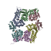
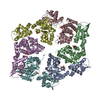
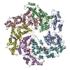
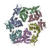
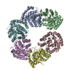
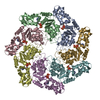
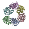
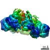
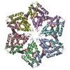
| Title | Crystal structure of symmetric W-W-W ClpX Hexamer | ||||||
 Components Components | ATP-dependent Clp protease ATP-binding subunit ClpX | ||||||
 Keywords Keywords | MOTOR PROTEIN / ATPase / symmetric / hexamer | ||||||
| Function / homology |  Function and homology information Function and homology informationHslUV protease complex / endopeptidase Clp complex / ATP-dependent peptidase activity / protein unfolding / : / ATP-dependent protein folding chaperone / disordered domain specific binding / : / protease binding / protein dimerization activity ...HslUV protease complex / endopeptidase Clp complex / ATP-dependent peptidase activity / protein unfolding / : / ATP-dependent protein folding chaperone / disordered domain specific binding / : / protease binding / protein dimerization activity / cell division / ATP hydrolysis activity / zinc ion binding / ATP binding / identical protein binding / cytosol Similarity search - Function | ||||||
| Biological species |  | ||||||
| Method |  X-RAY DIFFRACTION / SYNCHROTRON / MOLECULAR REPLACEMENT / Resolution: 5.0003 Å X-RAY DIFFRACTION / SYNCHROTRON / MOLECULAR REPLACEMENT / Resolution: 5.0003 Å | ||||||
 Authors Authors | Glynn, S.E. / Nager, A.R. / Stinson, B.S. / Schmitz, K.R. / Baker, T.A. / Sauer, R.T. | ||||||
 Citation Citation | Journal: Cell(Cambridge,Mass.) / Year: 2013 Title: Nucleotide Binding and Conformational Switching in the Hexameric Ring of a AAA+ Machine. Authors: Stinson, B.M. / Nager, A.R. / Glynn, S.E. / Schmitz, K.R. / Baker, T.A. / Sauer, R.T. | ||||||
| History |
|
- Structure visualization
| Structure viewer | Molecule: MolmilJmol/JSmol |
|---|
- Downloads & links
-Download
| PDBx/mmCIF format | 4i9k.cif.gz | 238.1 KB | Display | PDBx/mmCIF format |
|---|---|---|---|---|
| PDB format | pdb4i9k.ent.gz | 193.7 KB | Display | PDB format |
| PDBx/mmJSON format | 4i9k.json.gz | Tree view | PDBx/mmJSON format | |
| Others |  Other downloads Other downloads |
-Validation report
| Arichive directory | https://data.pdbj.org/pub/pdb/validation_reports/i9/4i9kftp://data.pdbj.org/pub/pdb/validation_reports/i9/4i9k | HTTPS FTP |
|---|
-Related structure data
| Related structure data | 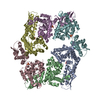 4i34C 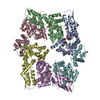 4i4lC  4i5oC  4i63C 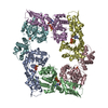 4i81C 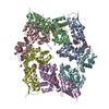 3hwsS S: Starting model for refinement C: citing same article ( |
|---|---|
| Similar structure data |
-Links
 PDBj
PDBj

- Assembly
| Deposited unit | 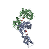
| ||||||||
|---|---|---|---|---|---|---|---|---|---|
| 1 | 
| ||||||||
| Unit cell |
|
-Components
| #1: Protein | Mass: 39435.793 Da / Num. of mol.: 2 Source method: isolated from a genetically manipulated source Source: (gene. exp.) #2: Chemical | ChemComp-SO4 /   Mass: 96.063 Da / Num. of mol.: 4 / Source method: obtained synthetically / Formula: SO4 Mass: 96.063 Da / Num. of mol.: 4 / Source method: obtained synthetically / Formula: SO4 |
|---|
-Experimental details
-Experiment
| Experiment | Method: X-RAY DIFFRACTION / Number of used crystals: 1 |
|---|
- Sample preparation
| Crystal | Density Matthews: 2.92 Å3/Da / Density % sol: 57.82 % |
|---|---|
| Crystal grow | Temperature: 298 K / Method: vapor diffusion / pH: 7.5 Details: 2 M ammonium sulfate, 0.15 M potassium sulfate, 4 mM ATP, 4 mM magnesium sulfate, 50 mM EDTA, pH 7.5, VAPOR DIFFUSION, temperature 298K |
-Data collection
| Diffraction | Mean temperature: 100 K | ||||||||||||||||||||||||||||||||||||||||||||||||||||||||||||||||||||||||||||||||||||||||
|---|---|---|---|---|---|---|---|---|---|---|---|---|---|---|---|---|---|---|---|---|---|---|---|---|---|---|---|---|---|---|---|---|---|---|---|---|---|---|---|---|---|---|---|---|---|---|---|---|---|---|---|---|---|---|---|---|---|---|---|---|---|---|---|---|---|---|---|---|---|---|---|---|---|---|---|---|---|---|---|---|---|---|---|---|---|---|---|---|---|
| Diffraction source | Source: SYNCHROTRON / Site: APS  / Beamline: 24-ID-C / Wavelength: 0.979 Å / Beamline: 24-ID-C / Wavelength: 0.979 Å | ||||||||||||||||||||||||||||||||||||||||||||||||||||||||||||||||||||||||||||||||||||||||
| Detector | Type: ADSC QUANTUM 315 / Detector: CCD / Date: Jun 1, 2010 | ||||||||||||||||||||||||||||||||||||||||||||||||||||||||||||||||||||||||||||||||||||||||
| Radiation | Protocol: SINGLE WAVELENGTH / Scattering type: x-ray | ||||||||||||||||||||||||||||||||||||||||||||||||||||||||||||||||||||||||||||||||||||||||
| Radiation wavelength | Wavelength: 0.979 Å / Relative weight: 1 | ||||||||||||||||||||||||||||||||||||||||||||||||||||||||||||||||||||||||||||||||||||||||
| Reflection | Resolution: 4.506→59.715 Å / Num. all: 5160 / Num. obs: 5160 / % possible obs: 94.6 % / Observed criterion σ(F): 2 / Observed criterion σ(I): 2 / Redundancy: 6.4 % / Rsym value: 0.186 / Net I/σ(I): 5.5 | ||||||||||||||||||||||||||||||||||||||||||||||||||||||||||||||||||||||||||||||||||||||||
| Reflection shell | Diffraction-ID: 1
|
- Processing
| Software |
| ||||||||||||||||||||||||||||
|---|---|---|---|---|---|---|---|---|---|---|---|---|---|---|---|---|---|---|---|---|---|---|---|---|---|---|---|---|---|
| Refinement | Method to determine structure: MOLECULAR REPLACEMENT Starting model: PDB ENTRY 3HWS Resolution: 5.0003→49.15 Å / Occupancy max: 1 / Occupancy min: 1 / FOM work R set: 0.5862 / SU ML: 0.75 / Phase error: 44.39 / Stereochemistry target values: ML
| ||||||||||||||||||||||||||||
| Solvent computation | Shrinkage radii: 1.2 Å / VDW probe radii: 1.4 Å / Solvent model: FLAT BULK SOLVENT MODEL / Bsol: 188.461 Å2 / ksol: 0.341 e/Å3 | ||||||||||||||||||||||||||||
| Displacement parameters | Biso max: 290.44 Å2 / Biso mean: 240.9206 Å2 / Biso min: 194.68 Å2
| ||||||||||||||||||||||||||||
| Refinement step | Cycle: LAST / Resolution: 5.0003→49.15 Å
| ||||||||||||||||||||||||||||
| Refine LS restraints |
| ||||||||||||||||||||||||||||
| LS refinement shell | Refine-ID: X-RAY DIFFRACTION / Total num. of bins used: 3
|
