 Movie
Movie Controller

[English] 日本語
 Yorodumi
- PDB-4i9c: Crystal structure of aspartyl phosphate phosphatase F from B.subt... -
+ Open data

- Basic information
| Entry | Database: PDB / ID: 4i9c | ||||||
|---|---|---|---|---|---|---|---|
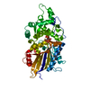
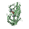
| Title | Crystal structure of aspartyl phosphate phosphatase F from B.subtilis in complex with its inhibitory peptide | ||||||
 Components Components |
| ||||||
 Keywords Keywords | GENE REGULATION / ComA inhibitor / PhrF | ||||||
| Function / homology |  Function and homology information Function and homology informationestablishment of competence for transformation / metal ion binding / cytoplasm Similarity search - Function | ||||||
| Biological species |  | ||||||
| Method |  X-RAY DIFFRACTION / SYNCHROTRON / SIRAS / Resolution: 3.1 Å X-RAY DIFFRACTION / SYNCHROTRON / SIRAS / Resolution: 3.1 Å | ||||||
 Authors Authors | Marina, A. / Gallego, F. | ||||||
 Citation Citation | Journal: Plos Biol. / Year: 2013 Title: Structural basis of Rap phosphatase inhibition by Phr peptides Authors: Gallego del Sol, F. / Marina, A. | ||||||
| History |
|
- Structure visualization
| Structure viewer | Molecule: MolmilJmol/JSmol |
|---|
- Downloads & links
-Download
| PDBx/mmCIF format | 4i9c.cif.gz | 171.3 KB | Display | PDBx/mmCIF format |
|---|---|---|---|---|
| PDB format | pdb4i9c.ent.gz | 139.9 KB | Display | PDB format |
| PDBx/mmJSON format | 4i9c.json.gz | Tree view | PDBx/mmJSON format | |
| Others |  Other downloads Other downloads |
-Validation report
| Arichive directory | https://data.pdbj.org/pub/pdb/validation_reports/i9/4i9cftp://data.pdbj.org/pub/pdb/validation_reports/i9/4i9c | HTTPS FTP |
|---|
-Related structure data


-Links
 PDBj
PDBj


- Assembly
| Deposited unit | 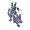
| ||||||||
|---|---|---|---|---|---|---|---|---|---|
| 1 |
| ||||||||
| Unit cell |
|
-Components
| #1: Protein | Mass: 45664.965 Da / Num. of mol.: 1 Source method: isolated from a genetically manipulated source Source: (gene. exp.) References: UniProt: P71002, Hydrolases; Acting on ester bonds |
|---|---|
| #2: Protein/peptide | Mass: 604.743 Da / Num. of mol.: 1 / Source method: obtained synthetically / Details: peptide purchased from Genescript |
| #3: Water | ChemComp-HOH /  Mass: 18.015 Da / Num. of mol.: 18 / Source method: isolated from a natural source / Formula: H2O Mass: 18.015 Da / Num. of mol.: 18 / Source method: isolated from a natural source / Formula: H2O |
-Experimental details
-Experiment
| Experiment | Method: X-RAY DIFFRACTION / Number of used crystals: 2 |
|---|
- Sample preparation
| Crystal | Density Matthews: 4.85 Å3/Da / Density % sol: 74.62 % |
|---|---|
| Crystal grow | Temperature: 294 K / Method: vapor diffusion, sitting drop / pH: 8.5 Details: 1M ammonium sulphate, 17% glycerol, 0.1M tris pH 8.5, VAPOR DIFFUSION, SITTING DROP, temperature 294K |
-Data collection
| Diffraction |
| |||||||||||||||
|---|---|---|---|---|---|---|---|---|---|---|---|---|---|---|---|---|
| Diffraction source |
| |||||||||||||||
| Detector |
| |||||||||||||||
| Radiation | Monochromator: Si(111) channel-cut / Protocol: SINGLE WAVELENGTH / Monochromatic (M) / Laue (L): M / Scattering type: x-ray | |||||||||||||||
| Radiation wavelength |
| |||||||||||||||
| Reflection | Resolution: 3.1→49.37 Å / Num. all: 16988 / Num. obs: 16991 / % possible obs: 100 % / Observed criterion σ(F): 1 / Observed criterion σ(I): 1 / Redundancy: 22 % / Biso Wilson estimate: 107.9 Å2 / Rmerge(I) obs: 0.097 / Rsym value: 0.095 / Net I/σ(I): 31.3 | |||||||||||||||
| Reflection shell | Resolution: 3.1→3.26 Å / Redundancy: 22.3 % / Rmerge(I) obs: 0.265 / Mean I/σ(I) obs: 3.3 / Num. unique all: 2427 / Rsym value: 0.265 / % possible all: 100 |


- Processing
| Software |
| |||||||||||||||||||||||||||||||||||||||||||||
|---|---|---|---|---|---|---|---|---|---|---|---|---|---|---|---|---|---|---|---|---|---|---|---|---|---|---|---|---|---|---|---|---|---|---|---|---|---|---|---|---|---|---|---|---|---|---|
| Refinement | Method to determine structure: SIRAS / Resolution: 3.1→47.07 Å / Cor.coef. Fo:Fc: 0.953 / Cor.coef. Fo:Fc free: 0.941 / SU B: 27.984 / SU ML: 0.243 / Cross valid method: THROUGHOUT / σ(F): 2 / ESU R: 0.639 / ESU R Free: 0.328 / Stereochemistry target values: MAXIMUM LIKELIHOOD / Details: HYDROGENS HAVE BEEN USED IF PRESENT IN THE INPUT
| |||||||||||||||||||||||||||||||||||||||||||||
| Solvent computation | Ion probe radii: 0.8 Å / Shrinkage radii: 0.8 Å / VDW probe radii: 1.2 Å / Solvent model: MASK | |||||||||||||||||||||||||||||||||||||||||||||
| Displacement parameters | Biso mean: 96.074 Å2 | |||||||||||||||||||||||||||||||||||||||||||||
| Refinement step | Cycle: LAST / Resolution: 3.1→47.07 Å
| |||||||||||||||||||||||||||||||||||||||||||||
| Refine LS restraints |
| |||||||||||||||||||||||||||||||||||||||||||||
| LS refinement shell | Resolution: 3.1→3.181 Å / Total num. of bins used: 20
| |||||||||||||||||||||||||||||||||||||||||||||
| Refinement TLS params. | Method: refined / Origin x: 8.129 Å / Origin y: 42.168 Å / Origin z: 2.106 Å
|
