 Movie
Movie Controller

[English] 日本語
 Yorodumi
- PDB-4i82: Crystal Structure of Hypothetical Thioesterase Protein SP_1851 fr... -
+ Open data

- Basic information
| Entry | Database: PDB / ID: 4i82 | ||||||
|---|---|---|---|---|---|---|---|
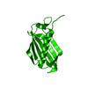
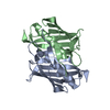
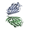
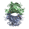
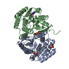
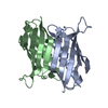
| Title | Crystal Structure of Hypothetical Thioesterase Protein SP_1851 from Streptococcus pneumoniae TIGR4 | ||||||
 Components Components | Putative uncharacterized protein | ||||||
 Keywords Keywords | HYDROLASE / PaaI/YdiI-like / Hot Dog Fold / thioesterase | ||||||
| Function / homology |  Function and homology information Function and homology information | ||||||
| Biological species |   Streptococcus pneumoniae (bacteria) Streptococcus pneumoniae (bacteria) | ||||||
| Method |  X-RAY DIFFRACTION / SYNCHROTRON / MOLECULAR REPLACEMENT / Resolution: 2.5 Å X-RAY DIFFRACTION / SYNCHROTRON / MOLECULAR REPLACEMENT / Resolution: 2.5 Å | ||||||
 Authors Authors | Khandokar, Y.B. / Forwood, J.K. | ||||||
 Citation Citation | Journal: To be published Title: Crystal Structure of Hypothetical Thioesterase Protein SP_1851 from Streptococcus pneumoniae TIGR4 Authors: Khandokar, Y.B. / Forwood, J.K. | ||||||
| History |
|
- Structure visualization
| Structure viewer | Molecule: MolmilJmol/JSmol |
|---|
- Downloads & links
-Download
| PDBx/mmCIF format | 4i82.cif.gz | 57.4 KB | Display | PDBx/mmCIF format |
|---|---|---|---|---|
| PDB format | pdb4i82.ent.gz | 42.4 KB | Display | PDB format |
| PDBx/mmJSON format | 4i82.json.gz | Tree view | PDBx/mmJSON format | |
| Others |  Other downloads Other downloads |
-Validation report
| Arichive directory | https://data.pdbj.org/pub/pdb/validation_reports/i8/4i82ftp://data.pdbj.org/pub/pdb/validation_reports/i8/4i82 | HTTPS FTP |
|---|
-Related structure data
| Related structure data |  4zrfC  3lbbS S: Starting model for refinement C: citing same article ( |
|---|---|
| Similar structure data |

-Links
 PDBj
PDBj
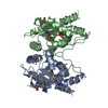
- Assembly
| Deposited unit | 
| ||||||||
|---|---|---|---|---|---|---|---|---|---|
| 1 | 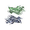
| ||||||||
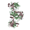
| Unit cell |
|
-Components
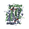
| #1: Protein | Mass: 15235.274 Da / Num. of mol.: 2 Source method: isolated from a genetically manipulated source Source: (gene. exp.) Streptococcus pneumoniae (bacteria) / Strain: TIGR4 / Gene: SP_1851 / Plasmid: pMCSG21 / Production host: |
|---|
-Experimental details
-Experiment
| Experiment | Method: X-RAY DIFFRACTION / Number of used crystals: 1 |
|---|
- Sample preparation
| Crystal | Density Matthews: 1.97 Å3/Da / Density % sol: 37.56 % |
|---|---|
| Crystal grow | Temperature: 290 K / Method: vapor diffusion, hanging drop / pH: 7.5 Details: 200mM Ammonium Tartrate dibasic, 20% PEG 3350, 100mM Tris pH7.5, VAPOR DIFFUSION, HANGING DROP, temperature 290K |
-Data collection
| Diffraction | Mean temperature: 100 K |
|---|---|
| Diffraction source | Source: SYNCHROTRON / Site: Australian Synchrotron  / Beamline: MX1 / Wavelength: 0.9537 Å / Beamline: MX1 / Wavelength: 0.9537 Å |
| Detector | Type: ADSC QUANTUM 315r / Detector: CCD / Date: Oct 9, 2012 |
| Radiation | Monochromator: Silicon Double Crystal / Protocol: SINGLE WAVELENGTH / Monochromatic (M) / Laue (L): M / Scattering type: x-ray |
| Radiation wavelength | Wavelength: 0.9537 Å / Relative weight: 1 |
| Reflection | Resolution: 2.49→35.22 Å / Num. all: 8714 / Num. obs: 8159 / % possible obs: 93.6 % / Observed criterion σ(F): 0 / Observed criterion σ(I): 0 |
| Reflection shell | Resolution: 2.49→2.62 Å / % possible all: 83.3 |
- Processing
| Software |
| ||||||||||||||||||||||||||||||||||||||||||||||||||||||||||||
|---|---|---|---|---|---|---|---|---|---|---|---|---|---|---|---|---|---|---|---|---|---|---|---|---|---|---|---|---|---|---|---|---|---|---|---|---|---|---|---|---|---|---|---|---|---|---|---|---|---|---|---|---|---|---|---|---|---|---|---|---|---|
| Refinement | Method to determine structure: MOLECULAR REPLACEMENT Starting model: 3LBB Resolution: 2.5→24.77 Å / Cor.coef. Fo:Fc: 0.911 / Cor.coef. Fo:Fc free: 0.877 / SU B: 12.567 / SU ML: 0.283 / Cross valid method: THROUGHOUT / ESU R: 1.174 / ESU R Free: 0.359 / Stereochemistry target values: MAXIMUM LIKELIHOOD / Details: HYDROGENS HAVE BEEN ADDED IN THE RIDING POSITIONS
| ||||||||||||||||||||||||||||||||||||||||||||||||||||||||||||
| Solvent computation | Ion probe radii: 0.8 Å / Shrinkage radii: 0.8 Å / VDW probe radii: 1.2 Å / Solvent model: MASK | ||||||||||||||||||||||||||||||||||||||||||||||||||||||||||||
| Displacement parameters | Biso mean: 26.826 Å2
| ||||||||||||||||||||||||||||||||||||||||||||||||||||||||||||
| Refinement step | Cycle: LAST / Resolution: 2.5→24.77 Å
| ||||||||||||||||||||||||||||||||||||||||||||||||||||||||||||
| Refine LS restraints |
| ||||||||||||||||||||||||||||||||||||||||||||||||||||||||||||
| LS refinement shell | Resolution: 2.5→2.564 Å / Total num. of bins used: 20
|
