 Movie
Movie Controller

[English] 日本語
 Yorodumi
- PDB-4i25: 2.00 Angstroms X-ray crystal structure of NAD- and substrate-boun... -
+ Open data

- Basic information
| Entry | Database: PDB / ID: 4i25 | ||||||
|---|---|---|---|---|---|---|---|
| Title | 2.00 Angstroms X-ray crystal structure of NAD- and substrate-bound 2-aminomuconate 6-semialdehyde dehydrogenase from Pseudomonas fluorescens | ||||||
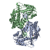
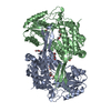
 Components Components | 2-aminomuconate 6-semialdehyde dehydrogenase | ||||||
 Keywords Keywords | OXIDOREDUCTASE / Aldehyde dehydrogenase / Dehydrogenase / NAD | ||||||
| Function / homology |  Function and homology information Function and homology informationoxidoreductase activity, acting on the aldehyde or oxo group of donors, NAD or NADP as acceptor / nucleotide binding Similarity search - Function | ||||||
| Biological species |  Pseudomonas fluorescens (bacteria) Pseudomonas fluorescens (bacteria) | ||||||
| Method |  X-RAY DIFFRACTION / SYNCHROTRON / MOLECULAR REPLACEMENT / Resolution: 2 Å X-RAY DIFFRACTION / SYNCHROTRON / MOLECULAR REPLACEMENT / Resolution: 2 Å | ||||||
 Authors Authors | Huo, L. / Davis, I. / Chen, L. / Liu, A. | ||||||
 Citation Citation | Journal: Nat Commun / Year: 2015 Title: Crystallographic and spectroscopic snapshots reveal a dehydrogenase in action. Authors: Huo, L. / Davis, I. / Liu, F. / Andi, B. / Esaki, S. / Iwaki, H. / Hasegawa, Y. / Orville, A.M. / Liu, A. | ||||||
| History |
|
- Structure visualization
| Structure viewer | Molecule: MolmilJmol/JSmol |
|---|
- Downloads & links
-Download
| PDBx/mmCIF format | 4i25.cif.gz | 404.4 KB | Display | PDBx/mmCIF format |
|---|---|---|---|---|
| PDB format | pdb4i25.ent.gz | 326.8 KB | Display | PDB format |
| PDBx/mmJSON format | 4i25.json.gz | Tree view | PDBx/mmJSON format | |
| Others |  Other downloads Other downloads |
-Validation report
| Arichive directory | https://data.pdbj.org/pub/pdb/validation_reports/i2/4i25ftp://data.pdbj.org/pub/pdb/validation_reports/i2/4i25 | HTTPS FTP |
|---|
-Related structure data
| Related structure data |  4i1wC  4i26C  4i2rC  4npiC  4oe2C  4ou2C  4oubC 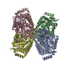 2d4eS C: citing same article ( S: Starting model for refinement |
|---|---|
| Similar structure data |


-Links
 PDBj
PDBj

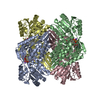
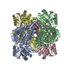
- Assembly
| Deposited unit | 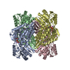
| ||||||||
|---|---|---|---|---|---|---|---|---|---|
| 1 |
| ||||||||
| Unit cell |
|
-Components
| #1: Protein | Mass: 53975.680 Da / Num. of mol.: 4 Source method: isolated from a genetically manipulated source Source: (gene. exp.) Pseudomonas fluorescens (bacteria) / Strain: KU-7 / Gene: nbaE / Plasmid: pET16Bb / Production host: #2: Chemical | ChemComp-NAD /   Mass: 663.425 Da / Num. of mol.: 4 / Source method: obtained synthetically / Formula: C21H27N7O14P2 / Comment: NAD*YM Mass: 663.425 Da / Num. of mol.: 4 / Source method: obtained synthetically / Formula: C21H27N7O14P2 / Comment: NAD*YM#3: Chemical | ChemComp-NA /   Mass: 22.990 Da / Num. of mol.: 4 / Source method: obtained synthetically / Formula: Na Mass: 22.990 Da / Num. of mol.: 4 / Source method: obtained synthetically / Formula: Na#4: Chemical | ChemComp-6OD / ( 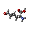  Mass: 141.125 Da / Num. of mol.: 4 / Source method: obtained synthetically / Formula: C6H7NO3 Mass: 141.125 Da / Num. of mol.: 4 / Source method: obtained synthetically / Formula: C6H7NO3#5: Water | ChemComp-HOH / |  Mass: 18.015 Da / Num. of mol.: 1397 / Source method: isolated from a natural source / Formula: H2O Mass: 18.015 Da / Num. of mol.: 1397 / Source method: isolated from a natural source / Formula: H2O |
|---|
-Experimental details
-Experiment
| Experiment | Method: X-RAY DIFFRACTION / Number of used crystals: 1 |
|---|
- Sample preparation
| Crystal | Density Matthews: 2.56 Å3/Da / Density % sol: 51.91 % |
|---|---|
| Crystal grow | Temperature: 291 K / Method: vapor diffusion, hanging drop / pH: 9.1 Details: 0.2 M Sodium phosphate dibasic dihydrate, 20% w/v Polyethylene glycol 3,350 , pH 9.1, VAPOR DIFFUSION, HANGING DROP, temperature 291K |
-Data collection
| Diffraction | Mean temperature: 100 K |
|---|---|
| Diffraction source | Source: SYNCHROTRON / Site: APS  / Beamline: 22-ID / Wavelength: 0.8 Å / Beamline: 22-ID / Wavelength: 0.8 Å |
| Detector | Type: MARMOSAIC 300 mm CCD / Detector: CCD / Date: Jul 22, 2012 |
| Radiation | Protocol: SINGLE WAVELENGTH / Monochromatic (M) / Laue (L): M / Scattering type: x-ray |
| Radiation wavelength | Wavelength: 0.8 Å / Relative weight: 1 |
| Reflection | Resolution: 2→35 Å / Num. all: 149047 / Num. obs: 149047 / % possible obs: 100 % / Observed criterion σ(F): 0 / Observed criterion σ(I): 0 / Redundancy: 14.4 % / Biso Wilson estimate: 26.67 Å2 / Rmerge(I) obs: 0.12 / Net I/σ(I): 34 |
| Reflection shell | Resolution: 2→2.03 Å / % possible all: 100 |
- Processing
| Software |
| ||||||||||||||||||||||||||||||||||||||||||||||||||||||||||||
|---|---|---|---|---|---|---|---|---|---|---|---|---|---|---|---|---|---|---|---|---|---|---|---|---|---|---|---|---|---|---|---|---|---|---|---|---|---|---|---|---|---|---|---|---|---|---|---|---|---|---|---|---|---|---|---|---|---|---|---|---|---|
| Refinement | Method to determine structure: MOLECULAR REPLACEMENT Starting model: PDB entry 2D4E Resolution: 2→27.22 Å / Cor.coef. Fo:Fc: 0.967 / Cor.coef. Fo:Fc free: 0.947 / Occupancy max: 1 / Occupancy min: 1 / SU B: 3.567 / SU ML: 0.099 / Cross valid method: THROUGHOUT / σ(F): 0 / ESU R: 0.155 / ESU R Free: 0.145 / Stereochemistry target values: MAXIMUM LIKELIHOOD Details: HYDROGENS HAVE BEEN ADDED IN THE RIDING POSITIONS U VALUES : REFINED INDIVIDUALLY
| ||||||||||||||||||||||||||||||||||||||||||||||||||||||||||||
| Solvent computation | Ion probe radii: 0.8 Å / Shrinkage radii: 0.8 Å / VDW probe radii: 1.2 Å / Solvent model: MASK | ||||||||||||||||||||||||||||||||||||||||||||||||||||||||||||
| Displacement parameters | Biso max: 87.93 Å2 / Biso mean: 29.3828 Å2 / Biso min: 13.27 Å2
| ||||||||||||||||||||||||||||||||||||||||||||||||||||||||||||
| Refinement step | Cycle: LAST / Resolution: 2→27.22 Å
| ||||||||||||||||||||||||||||||||||||||||||||||||||||||||||||
| Refine LS restraints |
| ||||||||||||||||||||||||||||||||||||||||||||||||||||||||||||
| LS refinement shell | Resolution: 2.001→2.053 Å / Total num. of bins used: 20
|
