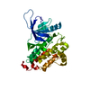
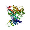
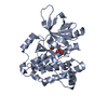
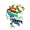
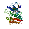
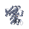
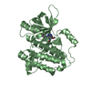
Entry Database : PDB / ID : 4i24Title Structure of T790M EGFR kinase domain co-crystallized with dacomitinib Epidermal growth factor receptor Keywords / / / Function / homology Function Domain/homology Component
/ / / / / / / / / / / / / / / / / / / / / / / / / / / / / / / / / / / / / / / / / / / / / / / / / / / / / / / / / / / / / / / / / / / / / / / / / / / / / / / / / / / / / / / / / / / / / / / / / / / / / / / / / / / / / / / / / / / / / / / / / / / / / / / / / / / / / / / / / / / / / / / / / / / / / Biological species Homo sapiens (human)Method / / / Resolution : 1.8 Å Authors Gajiwala, K.S. / Feng, J. / Ferre, R. / Ryan, K. / Brodsky, O. / Stewart, A. Journal : Structure / Year : 2013Title : Insights into the Aberrant Activity of Mutant EGFR Kinase Domain and Drug Recognition.Authors : Gajiwala, K.S. / Feng, J. / Ferre, R. / Ryan, K. / Brodsky, O. / Weinrich, S. / Kath, J.C. / Stewart, A. History Deposition Nov 21, 2012 Deposition site / Processing site Revision 1.0 Jan 16, 2013 Provider / Type Revision 1.1 Feb 27, 2013 Group Revision 1.2 Nov 27, 2024 Group Data collection / Database references ... Data collection / Database references / Derived calculations / Structure summary Category chem_comp_atom / chem_comp_bond ... chem_comp_atom / chem_comp_bond / database_2 / pdbx_entry_details / pdbx_modification_feature / struct_conn / struct_ref_seq_dif / struct_site Item _database_2.pdbx_DOI / _database_2.pdbx_database_accession ... _database_2.pdbx_DOI / _database_2.pdbx_database_accession / _struct_conn.pdbx_dist_value / _struct_conn.pdbx_leaving_atom_flag / _struct_conn.ptnr1_auth_asym_id / _struct_conn.ptnr1_label_asym_id / _struct_conn.ptnr2_auth_asym_id / _struct_conn.ptnr2_label_asym_id / _struct_ref_seq_dif.details / _struct_site.pdbx_auth_asym_id / _struct_site.pdbx_auth_comp_id / _struct_site.pdbx_auth_seq_id
Show all Show less
 Movie
Movie Controller

 Yorodumi
 Open data
 Basic information
 Components
 Keywords
 Function and homology information
 Homo sapiens (human)
 X-RAY DIFFRACTION /
  Authors
 Citation
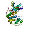
 Structure visualization
 Downloads & links
 Other downloads
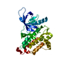


 PDBj
PDBj


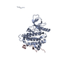
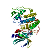
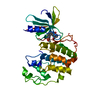
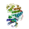
 Assembly
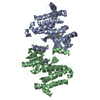

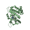
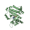
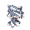
 Spodoptera frugiperda (fall armyworm)
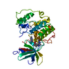
 Mass: 469.939 Da / Num. of mol.: 2 / Source method: obtained synthetically / Formula: C24H25ClFN5O2 / Comment: medication, inhibitor*YM
 Mass: 18.015 Da / Num. of mol.: 282 / Source method: isolated from a natural source / Formula: H2O
 Sample preparation
 / Beamline: 5.0.1 / Wavelength: 1 Å
 Processing