 Movie
Movie Controller

[English] 日本語
 Yorodumi
- PDB-4hzp: The Structure of the Bifunctional Acetyltransferase/Decarboxylase... -
+ Open data

- Basic information
| Entry | Database: PDB / ID: 4hzp | ||||||
|---|---|---|---|---|---|---|---|
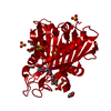
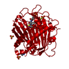
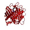
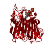
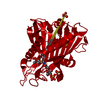
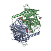
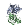
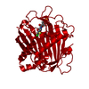
| Title | The Structure of the Bifunctional Acetyltransferase/Decarboxylase LnmK from the Leinamycin Biosynthetic Pathway Revealing Novel Activity for a Double Hot Dog Fold | ||||||
 Components Components | Bifunctional methylmalonyl-CoA:ACP Acyltransferase/Decarboxylase | ||||||
 Keywords Keywords | TRANSFERASE / LYASE / Structural Genomics / Protein Structure Initiative / Enzyme Discovery for Natural Product Biosynthesis / NatPro / PSI-Biology / Double Hot Dog Fold / Acyl Carrier Protein - LnmL / methylmalonyl-CoA | ||||||
| Function / homology |  Function and homology information Function and homology information | ||||||
| Biological species |  Streptomyces atroolivaceus (bacteria) Streptomyces atroolivaceus (bacteria) | ||||||
| Method |  X-RAY DIFFRACTION / SYNCHROTRON / MOLECULAR REPLACEMENT / Resolution: 1.77 Å X-RAY DIFFRACTION / SYNCHROTRON / MOLECULAR REPLACEMENT / Resolution: 1.77 Å | ||||||
 Authors Authors | Lohman, J.R. / Bingman, C.A. / Phillips Jr., G.N. / Shen, B. / Enzyme Discovery for Natural Product Biosynthesis (NatPro) | ||||||
 Citation Citation | Journal: Biochemistry / Year: 2013 Title: Structure of the Bifunctional Acyltransferase/Decarboxylase LnmK from the Leinamycin Biosynthetic Pathway Revealing Novel Activity for a Double-Hot-Dog Fold. Authors: Lohman, J.R. / Bingman, C.A. / Phillips, G.N. / Shen, B. | ||||||
| History |
|
- Structure visualization
| Structure viewer | Molecule: MolmilJmol/JSmol |
|---|
- Downloads & links
-Download
| PDBx/mmCIF format | 4hzp.cif.gz | 77.4 KB | Display | PDBx/mmCIF format |
|---|---|---|---|---|
| PDB format | pdb4hzp.ent.gz | 56.7 KB | Display | PDB format |
| PDBx/mmJSON format | 4hzp.json.gz | Tree view | PDBx/mmJSON format | |
| Others |  Other downloads Other downloads |
-Validation report
| Arichive directory | https://data.pdbj.org/pub/pdb/validation_reports/hz/4hzpftp://data.pdbj.org/pub/pdb/validation_reports/hz/4hzp | HTTPS FTP |
|---|
-Related structure data
| Related structure data | 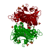 4hznSC  4hzoC S: Starting model for refinement C: citing same article ( |
|---|---|
| Similar structure data | |
| Other databases |


-Links
 PDBj
PDBj


- Assembly
| Deposited unit | 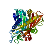
| ||||||||
|---|---|---|---|---|---|---|---|---|---|
| 1 | 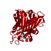
| ||||||||
| Unit cell |
| ||||||||
| Components on special symmetry positions |
| ||||||||
| Details | The biological dimer is generated by the symmetry mate: x, x-y, -z+1/6 |
-Components
| #1: Protein | Mass: 35263.285 Da / Num. of mol.: 1 / Mutation: T2S, Y62F Source method: isolated from a genetically manipulated source Source: (gene. exp.) Streptomyces atroolivaceus (bacteria) / Gene: LnmK / Plasmid: pETDuet-1 / Production host: | ||
|---|---|---|---|
| #2: Chemical | ChemComp-COA /   Mass: 767.534 Da / Num. of mol.: 1 / Source method: obtained synthetically / Formula: C21H36N7O16P3S Mass: 767.534 Da / Num. of mol.: 1 / Source method: obtained synthetically / Formula: C21H36N7O16P3S | ||
| #3: Chemical | ChemComp-CL /   Mass: 35.453 Da / Num. of mol.: 1 / Source method: obtained synthetically / Formula: Cl Mass: 35.453 Da / Num. of mol.: 1 / Source method: obtained synthetically / Formula: Cl | ||
| #4: Chemical |   Mass: 96.063 Da / Num. of mol.: 2 / Source method: obtained synthetically / Formula: SO4 Mass: 96.063 Da / Num. of mol.: 2 / Source method: obtained synthetically / Formula: SO4#5: Water | ChemComp-HOH / |  Mass: 18.015 Da / Num. of mol.: 160 / Source method: isolated from a natural source / Formula: H2O Mass: 18.015 Da / Num. of mol.: 160 / Source method: isolated from a natural source / Formula: H2O |
-Experimental details
-Experiment
| Experiment | Method: X-RAY DIFFRACTION / Number of used crystals: 1 |
|---|
- Sample preparation
| Crystal | Density Matthews: 2.27 Å3/Da / Density % sol: 45.82 % |
|---|---|
| Crystal grow | Temperature: 298 K / Method: vapor diffusion, hanging drop / pH: 7 Details: Protein: 20 mg/mL in 100 mM NaCl and 10 mM Tris-HCl, pH 8.0 plus 10 mM methylmalonyl-CoA. Precipitant: 20 - 23% MEPEG 2000, 0.25-0.35 M (NH4)2SO4 and 0.1 M Bis-tris propane-HCl, pH 7.0 in 4 ...Details: Protein: 20 mg/mL in 100 mM NaCl and 10 mM Tris-HCl, pH 8.0 plus 10 mM methylmalonyl-CoA. Precipitant: 20 - 23% MEPEG 2000, 0.25-0.35 M (NH4)2SO4 and 0.1 M Bis-tris propane-HCl, pH 7.0 in 4 L drops (1:1, protein:well). , VAPOR DIFFUSION, HANGING DROP, temperature 298K |
-Data collection
| Diffraction | Mean temperature: 100 K |
|---|---|
| Diffraction source | Source: SYNCHROTRON / Site: APS  / Beamline: 21-ID-G / Wavelength: 0.97857 Å / Beamline: 21-ID-G / Wavelength: 0.97857 Å |
| Detector | Type: MAR scanner 300 mm plate / Detector: IMAGE PLATE / Date: Dec 10, 2009 |
| Radiation | Monochromator: Si(111) / Protocol: SINGLE WAVELENGTH / Monochromatic (M) / Laue (L): M / Scattering type: x-ray |
| Radiation wavelength | Wavelength: 0.97857 Å / Relative weight: 1 |
| Reflection | Resolution: 1.77→30 Å / Num. all: 33385 / Num. obs: 32885 / % possible obs: 98.5 % / Observed criterion σ(I): -3 / Redundancy: 9.1 % / Rmerge(I) obs: 0.107 / Net I/σ(I): 14.7 |
- Processing
| Software |
| ||||||||||||||||||||||||||||||||||||||||||||||||||||||||||||||||||||||||||||||||||||||||||||||||||||||||||||||||||||||||||||||||||||||||||||||||||||||||||||||||||||||||||
|---|---|---|---|---|---|---|---|---|---|---|---|---|---|---|---|---|---|---|---|---|---|---|---|---|---|---|---|---|---|---|---|---|---|---|---|---|---|---|---|---|---|---|---|---|---|---|---|---|---|---|---|---|---|---|---|---|---|---|---|---|---|---|---|---|---|---|---|---|---|---|---|---|---|---|---|---|---|---|---|---|---|---|---|---|---|---|---|---|---|---|---|---|---|---|---|---|---|---|---|---|---|---|---|---|---|---|---|---|---|---|---|---|---|---|---|---|---|---|---|---|---|---|---|---|---|---|---|---|---|---|---|---|---|---|---|---|---|---|---|---|---|---|---|---|---|---|---|---|---|---|---|---|---|---|---|---|---|---|---|---|---|---|---|---|---|---|---|---|---|---|---|
| Refinement | Method to determine structure: MOLECULAR REPLACEMENT Starting model: PDB Entry 4HZN Resolution: 1.77→29.72 Å / Cor.coef. Fo:Fc: 0.937 / Cor.coef. Fo:Fc free: 0.904 / SU B: 3.366 / SU ML: 0.106 / Cross valid method: THROUGHOUT / ESU R: 0.142 / ESU R Free: 0.143 / Stereochemistry target values: MAXIMUM LIKELIHOOD / Details: HYDROGENS HAVE BEEN ADDED IN THE RIDING POSITIONS
| ||||||||||||||||||||||||||||||||||||||||||||||||||||||||||||||||||||||||||||||||||||||||||||||||||||||||||||||||||||||||||||||||||||||||||||||||||||||||||||||||||||||||||
| Solvent computation | Ion probe radii: 0.8 Å / Shrinkage radii: 0.8 Å / VDW probe radii: 1.4 Å / Solvent model: MASK | ||||||||||||||||||||||||||||||||||||||||||||||||||||||||||||||||||||||||||||||||||||||||||||||||||||||||||||||||||||||||||||||||||||||||||||||||||||||||||||||||||||||||||
| Displacement parameters | Biso mean: 27.185 Å2
| ||||||||||||||||||||||||||||||||||||||||||||||||||||||||||||||||||||||||||||||||||||||||||||||||||||||||||||||||||||||||||||||||||||||||||||||||||||||||||||||||||||||||||
| Refinement step | Cycle: LAST / Resolution: 1.77→29.72 Å
| ||||||||||||||||||||||||||||||||||||||||||||||||||||||||||||||||||||||||||||||||||||||||||||||||||||||||||||||||||||||||||||||||||||||||||||||||||||||||||||||||||||||||||
| Refine LS restraints |
| ||||||||||||||||||||||||||||||||||||||||||||||||||||||||||||||||||||||||||||||||||||||||||||||||||||||||||||||||||||||||||||||||||||||||||||||||||||||||||||||||||||||||||
| LS refinement shell | Resolution: 1.773→1.818 Å / Total num. of bins used: 20
|
