 Movie
Movie Controller

[English] 日本語
 Yorodumi
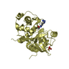
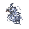
- PDB-4hyq: Crystal structure of phospholipase A1 from Streptomyces albidofla... -
+ Open data

- Basic information
| Entry | Database: PDB / ID: 4hyq | ||||||
|---|---|---|---|---|---|---|---|
| Title | Crystal structure of phospholipase A1 from Streptomyces albidoflavus NA297 | ||||||
 Components Components | phospholipase A1 | ||||||
 Keywords Keywords | HYDROLASE / lipase / glycerophospholipid | ||||||
| Function / homology |  Function and homology information Function and homology informationtriglyceride catabolic process / triacylglycerol lipase / triacylglycerol lipase activity Similarity search - Function | ||||||
| Biological species |  Streptomyces albidoflavus (bacteria) Streptomyces albidoflavus (bacteria) | ||||||
| Method |  X-RAY DIFFRACTION / SYNCHROTRON / MOLECULAR REPLACEMENT / Resolution: 1.75 Å X-RAY DIFFRACTION / SYNCHROTRON / MOLECULAR REPLACEMENT / Resolution: 1.75 Å | ||||||
 Authors Authors | Murayama, K. / Sugimori, D. | ||||||
 Citation Citation | Journal: J.Struct.Biol. / Year: 2013 Title: Crystal structure of phospholipase A1 from Streptomyces albidoflavus NA297 Authors: Murayama, K. / Kano, K. / Matsumoto, Y. / Sugimori, D. | ||||||
| History |
|
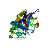
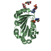
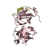
- Structure visualization
| Structure viewer | Molecule: MolmilJmol/JSmol |
|---|
- Downloads & links
-Download
| PDBx/mmCIF format | 4hyq.cif.gz | 59.6 KB | Display | PDBx/mmCIF format |
|---|---|---|---|---|
| PDB format | pdb4hyq.ent.gz | 43 KB | Display | PDB format |
| PDBx/mmJSON format | 4hyq.json.gz | Tree view | PDBx/mmJSON format | |
| Others |  Other downloads Other downloads |
-Validation report
| Summary document | 4hyq_validation.pdf.gz | 431.2 KB | Display | wwPDB validaton report |
|---|---|---|---|---|
| Full document | 4hyq_full_validation.pdf.gz | 432.9 KB | Display | |
| Data in XML | 4hyq_validation.xml.gz | 12.9 KB | Display | |
| Data in CIF | 4hyq_validation.cif.gz | 18.7 KB | Display | |
| Arichive directory | https://data.pdbj.org/pub/pdb/validation_reports/hy/4hyqftp://data.pdbj.org/pub/pdb/validation_reports/hy/4hyq | HTTPS FTP |
-Related structure data
| Related structure data |  1escS S: Starting model for refinement |
|---|---|
| Similar structure data |


-Links
 PDBj
PDBj- Assembly
| Deposited unit | 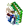
| ||||||||
|---|---|---|---|---|---|---|---|---|---|
| 1 |
| ||||||||
| Unit cell |
|
-Components
| #1: Protein | Mass: 24199.812 Da / Num. of mol.: 1 Source method: isolated from a genetically manipulated source Source: (gene. exp.) Streptomyces albidoflavus (bacteria) / Strain: NA297 / Plasmid: pUC702/pla / Production host: Streptomyces lividans (bacteria) / References: UniProt: K0J3J2*PLUS, phospholipase A1 |
|---|---|
| #2: Chemical | ChemComp-1PE /   Mass: 238.278 Da / Num. of mol.: 1 / Source method: obtained synthetically / Formula: C10H22O6 / Comment: precipitant*YM Mass: 238.278 Da / Num. of mol.: 1 / Source method: obtained synthetically / Formula: C10H22O6 / Comment: precipitant*YM |
| #3: Water | ChemComp-HOH /  Mass: 18.015 Da / Num. of mol.: 218 / Source method: isolated from a natural source / Formula: H2O Mass: 18.015 Da / Num. of mol.: 218 / Source method: isolated from a natural source / Formula: H2O |
| Has protein modification | Y |
| Sequence details | THE UNP SEQUENCE DATABASE REFERENCES FOR THIS PROTEIN DOES NOT CURRENTLY EXIST. THIS SEQUENCE HAS ...THE UNP SEQUENCE DATABASE REFERENCES |
-Experimental details
-Experiment
| Experiment | Method: X-RAY DIFFRACTION / Number of used crystals: 1 |
|---|
- Sample preparation
| Crystal | Density Matthews: 2.62 Å3/Da / Density % sol: 52.98 % |
|---|---|
| Crystal grow | Temperature: 293 K / Method: vapor diffusion, hanging drop / pH: 7.5 Details: 1.8M ammonium sulfate, 3% PEGMME2000, 0.1M Hepes, pH 7.5, VAPOR DIFFUSION, HANGING DROP, temperature 293K |
-Data collection
| Diffraction | Mean temperature: 100 K |
|---|---|
| Diffraction source | Source: SYNCHROTRON / Site: Photon Factory  / Beamline: BL-1A / Wavelength: 1 Å / Beamline: BL-1A / Wavelength: 1 Å |
| Detector | Type: ADSC QUANTUM 270 / Detector: CCD / Date: Mar 5, 2011 |
| Radiation | Monochromator: Si 111 / Protocol: SINGLE WAVELENGTH / Monochromatic (M) / Laue (L): M / Scattering type: x-ray |
| Radiation wavelength | Wavelength: 1 Å / Relative weight: 1 |
| Reflection | Resolution: 1.75→37.1 Å / Num. obs: 26008 / % possible obs: 100 % / Observed criterion σ(F): -3 / Redundancy: 10.9 % / Biso Wilson estimate: 14.9 Å2 / Rsym value: 0.105 / Net I/σ(I): 29.5 |
| Reflection shell | Resolution: 1.75→1.81 Å / Mean I/σ(I) obs: 5.8 / Rsym value: 0.618 / % possible all: 100 |
- Processing
| Software |
| ||||||||||||||||||||||||||||||||||||
|---|---|---|---|---|---|---|---|---|---|---|---|---|---|---|---|---|---|---|---|---|---|---|---|---|---|---|---|---|---|---|---|---|---|---|---|---|---|
| Refinement | Method to determine structure: MOLECULAR REPLACEMENT Starting model: PDB ENTRY 1ESC Resolution: 1.75→37.1 Å / Rfactor Rfree error: 0.006 / Data cutoff high absF: 1591373 / Data cutoff low absF: 0 / Isotropic thermal model: RESTRAINED / Cross valid method: THROUGHOUT / σ(F): 0 / Stereochemistry target values: Engh & Huber
| ||||||||||||||||||||||||||||||||||||
| Solvent computation | Solvent model: FLAT MODEL / Bsol: 61.3638 Å2 / ksol: 0.386993 e/Å3 | ||||||||||||||||||||||||||||||||||||
| Displacement parameters | Biso mean: 18.4 Å2
| ||||||||||||||||||||||||||||||||||||
| Refine analyze |
| ||||||||||||||||||||||||||||||||||||
| Refinement step | Cycle: LAST / Resolution: 1.75→37.1 Å
| ||||||||||||||||||||||||||||||||||||
| Refine LS restraints |
| ||||||||||||||||||||||||||||||||||||
| LS refinement shell | Resolution: 1.75→1.86 Å / Rfactor Rfree error: 0.018 / Total num. of bins used: 6
| ||||||||||||||||||||||||||||||||||||
| Xplor file |
|
