 Movie
Movie Controller

[English] 日本語
 Yorodumi
- PDB-4hyd: Structure of a presenilin family intramembrane aspartate protease... -
+ Open data

- Basic information
| Entry | Database: PDB / ID: 4hyd | ||||||
|---|---|---|---|---|---|---|---|
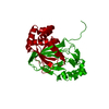
| Title | Structure of a presenilin family intramembrane aspartate protease in C2221 space group | ||||||
 Components Components | Putative uncharacterized protein | ||||||
 Keywords Keywords | MEMBRANE PROTEIN / protease | ||||||
| Function / homology | Signal-peptide peptidase, presenilin aspartyl protease / Signal-peptide peptidase, presenilin aspartyl protease / Presenilin/signal peptide peptidase / Presenilin, signal peptide peptidase, family / aspartic endopeptidase activity, intramembrane cleaving / endomembrane system / membrane / identical protein binding / Signal peptide peptidase Function and homology information Function and homology information | ||||||
| Biological species |  Methanoculleus marisnigri JR1 (archaea) Methanoculleus marisnigri JR1 (archaea) | ||||||
| Method |  X-RAY DIFFRACTION / SYNCHROTRON / MAD / Resolution: 3.8 Å X-RAY DIFFRACTION / SYNCHROTRON / MAD / Resolution: 3.8 Å | ||||||
 Authors Authors | Li, X. / Dang, S. / Yan, C. / Wang, J. / Shi, Y. | ||||||
 Citation Citation | Journal: Nature / Year: 2013 Title: Structure of a presenilin family intramembrane aspartate protease Authors: Li, X. / Dang, S. / Yan, C. / Gong, X. / Wang, J. / Shi, Y. | ||||||
| History |
|
- Structure visualization
| Structure viewer | Molecule: MolmilJmol/JSmol |
|---|
- Downloads & links
-Download
| PDBx/mmCIF format | 4hyd.cif.gz | 375.7 KB | Display | PDBx/mmCIF format |
|---|---|---|---|---|
| PDB format | pdb4hyd.ent.gz | 316.7 KB | Display | PDB format |
| PDBx/mmJSON format | 4hyd.json.gz | Tree view | PDBx/mmJSON format | |
| Others |  Other downloads Other downloads |
-Validation report
| Arichive directory | https://data.pdbj.org/pub/pdb/validation_reports/hy/4hydftp://data.pdbj.org/pub/pdb/validation_reports/hy/4hyd | HTTPS FTP |
|---|
-Related structure data


-Links
 PDBj
PDBj- Assembly
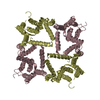
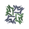
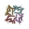
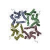
| Deposited unit | 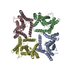
| ||||||||
|---|---|---|---|---|---|---|---|---|---|
| 1 |
| ||||||||
| Unit cell |
|
-Components
| #1: Protein | Mass: 31879.260 Da / Num. of mol.: 4 / Mutation: D40N, E42S, A147E, V148P, A229V Source method: isolated from a genetically manipulated source Source: (gene. exp.) Methanoculleus marisnigri JR1 (archaea)Strain: ATCC 35101 / DSM 1498 / JR1 / Gene: Memar_1924 / Production host:  |
|---|
-Experimental details
-Experiment
| Experiment | Method: X-RAY DIFFRACTION / Number of used crystals: 1 |
|---|
- Sample preparation
| Crystal | Density Matthews: 3.45 Å3/Da / Density % sol: 64.34 % |
|---|---|
| Crystal grow | Temperature: 291 K / Method: vapor diffusion, hanging drop / pH: 3.6 Details: 0.1M Glycine, 0.2M (NH4)2SO4, 20%(w/v) PEG500MME, 6%(w/v) Glycerol, 0.04%(w/v) Anapoe-C12E8, pH 3.6, VAPOR DIFFUSION, HANGING DROP, temperature 291K |
-Data collection
| Diffraction | Mean temperature: 100 K | ||||||||||||
|---|---|---|---|---|---|---|---|---|---|---|---|---|---|
| Diffraction source | Source: SYNCHROTRON / Site: SPring-8  / Beamline: BL41XU / Wavelength: 1.07171, 1.05384, 1.07206 / Beamline: BL41XU / Wavelength: 1.07171, 1.05384, 1.07206 | ||||||||||||
| Detector | Type: MARMOSAIC 225 mm CCD / Detector: CCD / Date: Apr 12, 2011 | ||||||||||||
| Radiation | Protocol: MAD / Monochromatic (M) / Laue (L): M / Scattering type: x-ray | ||||||||||||
| Radiation wavelength |
| ||||||||||||
| Reflection | Resolution: 3.8→50 Å / Num. obs: 15330 |
- Processing
| Software | Name: PHENIX / Version: (phenix.refine: 1.8_1069) / Classification: refinement | |||||||||||||||||||||||||||||||||||||||||||||||||||||||||||||||||||||||||||||||||||||||||||||||||||||||||||||||||||||||||||||
|---|---|---|---|---|---|---|---|---|---|---|---|---|---|---|---|---|---|---|---|---|---|---|---|---|---|---|---|---|---|---|---|---|---|---|---|---|---|---|---|---|---|---|---|---|---|---|---|---|---|---|---|---|---|---|---|---|---|---|---|---|---|---|---|---|---|---|---|---|---|---|---|---|---|---|---|---|---|---|---|---|---|---|---|---|---|---|---|---|---|---|---|---|---|---|---|---|---|---|---|---|---|---|---|---|---|---|---|---|---|---|---|---|---|---|---|---|---|---|---|---|---|---|---|---|---|---|
| Refinement | Method to determine structure: MAD / Resolution: 3.8→31.577 Å / SU ML: 0.55 / σ(F): 1.34 / Phase error: 53.18 / Stereochemistry target values: MLHL
| |||||||||||||||||||||||||||||||||||||||||||||||||||||||||||||||||||||||||||||||||||||||||||||||||||||||||||||||||||||||||||||
| Solvent computation | Shrinkage radii: 0.9 Å / VDW probe radii: 1.11 Å / Solvent model: FLAT BULK SOLVENT MODEL | |||||||||||||||||||||||||||||||||||||||||||||||||||||||||||||||||||||||||||||||||||||||||||||||||||||||||||||||||||||||||||||
| Refinement step | Cycle: LAST / Resolution: 3.8→31.577 Å
| |||||||||||||||||||||||||||||||||||||||||||||||||||||||||||||||||||||||||||||||||||||||||||||||||||||||||||||||||||||||||||||
| Refine LS restraints |
| |||||||||||||||||||||||||||||||||||||||||||||||||||||||||||||||||||||||||||||||||||||||||||||||||||||||||||||||||||||||||||||
| LS refinement shell | Refine-ID: X-RAY DIFFRACTION / Rfactor Rfree: 0 / Total num. of bins used: 5
| |||||||||||||||||||||||||||||||||||||||||||||||||||||||||||||||||||||||||||||||||||||||||||||||||||||||||||||||||||||||||||||
| Refinement TLS params. | Method: refined / Refine-ID: X-RAY DIFFRACTION
| |||||||||||||||||||||||||||||||||||||||||||||||||||||||||||||||||||||||||||||||||||||||||||||||||||||||||||||||||||||||||||||
| Refinement TLS group |
|
