 Movie
Movie Controller


+ Open data

- Basic information
| Entry | Database: PDB / ID: 4hwx | ||||||
|---|---|---|---|---|---|---|---|
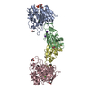
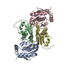
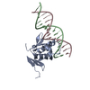
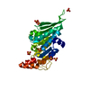
| Title | Crystal structure of Streptomyces caespitosus sermetstatin | ||||||
 Components Components | Neutral proteinase inhibitor ScNPI | ||||||
 Keywords Keywords | Hydrolase Inhibitor / Streptomyces subtilisin inhibitor fold | ||||||
| Function / homology |  Function and homology information Function and homology informationserine-type endopeptidase inhibitor activity / extracellular region / metal ion binding Similarity search - Function | ||||||
| Biological species |  Streptomyces caespitosus (bacteria) Streptomyces caespitosus (bacteria) | ||||||
| Method |  X-RAY DIFFRACTION / SYNCHROTRON / SAD / Resolution: 1.9 Å X-RAY DIFFRACTION / SYNCHROTRON / SAD / Resolution: 1.9 Å | ||||||
 Authors Authors | Trillo-Muyo, S. / Martinez-Rodriguez, S. / Arolas, J.L. / Gomis-Ruth, F.X. | ||||||
 Citation Citation | Journal: CHEM SCI / Year: 2013 Title: Mechanism of action of a Janus-faced single-domain protein inhibitor simultaneously targeting two peptidase classes Authors: Trillo-Muyo, S. / Martinez-Rodriguez, S. / Arolas, J.L. / Gomis-Ruth, F.X. | ||||||
| History |
|
- Structure visualization
| Structure viewer | Molecule: MolmilJmol/JSmol |
|---|
- Downloads & links
-Download
| PDBx/mmCIF format | 4hwx.cif.gz | 58.9 KB | Display | PDBx/mmCIF format |
|---|---|---|---|---|
| PDB format | pdb4hwx.ent.gz | 42.6 KB | Display | PDB format |
| PDBx/mmJSON format | 4hwx.json.gz | Tree view | PDBx/mmJSON format | |
| Others |  Other downloads Other downloads |
-Validation report
| Summary document | 4hwx_validation.pdf.gz | 440 KB | Display | wwPDB validaton report |
|---|---|---|---|---|
| Full document | 4hwx_full_validation.pdf.gz | 440.1 KB | Display | |
| Data in XML | 4hwx_validation.xml.gz | 9 KB | Display | |
| Data in CIF | 4hwx_validation.cif.gz | 11.7 KB | Display | |
| Arichive directory | https://data.pdbj.org/pub/pdb/validation_reports/hw/4hwxftp://data.pdbj.org/pub/pdb/validation_reports/hw/4hwx | HTTPS FTP |
-Related structure data


-Links
 PDBj
PDBj- Assembly
| Deposited unit | 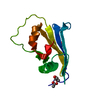
| ||||||||
|---|---|---|---|---|---|---|---|---|---|
| 1 | 
| ||||||||
| 2 | 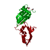
| ||||||||
| Unit cell |
|
-Components
| #1: Protein | Mass: 11923.354 Da / Num. of mol.: 1 Source method: isolated from a genetically manipulated source Source: (gene. exp.) Streptomyces caespitosus (bacteria) / Gene: ScNPI / Plasmid: pET32a / Production host: | ||||
|---|---|---|---|---|---|
| #2: Chemical | ChemComp-GOL /   Mass: 92.094 Da / Num. of mol.: 1 / Source method: obtained synthetically / Formula: C3H8O3 Mass: 92.094 Da / Num. of mol.: 1 / Source method: obtained synthetically / Formula: C3H8O3 | ||||
| #3: Chemical |   Mass: 59.044 Da / Num. of mol.: 3 / Source method: obtained synthetically / Formula: C2H3O2 Mass: 59.044 Da / Num. of mol.: 3 / Source method: obtained synthetically / Formula: C2H3O2#4: Water | ChemComp-HOH / |  Mass: 18.015 Da / Num. of mol.: 126 / Source method: isolated from a natural source / Formula: H2O Mass: 18.015 Da / Num. of mol.: 126 / Source method: isolated from a natural source / Formula: H2OHas protein modification | Y | |
-Experimental details
-Experiment
| Experiment | Method: X-RAY DIFFRACTION / Number of used crystals: 1 |
|---|
- Sample preparation
| Crystal | Density Matthews: 3.2 Å3/Da / Density % sol: 61.58 % |
|---|---|
| Crystal grow | Temperature: 293 K / Method: vapor diffusion, sitting drop / pH: 5.6 Details: 0.1 M sodium citrate dihydrate, 0.2 M ammonium acetate, 10% (w/v) PEG3350, pH 5.6, VAPOR DIFFUSION, SITTING DROP, temperature 293K |
-Data collection
| Diffraction | Mean temperature: 100 K |
|---|---|
| Diffraction source | Source: SYNCHROTRON / Site: ESRF  / Beamline: ID23-2 / Wavelength: 0.8726 Å / Beamline: ID23-2 / Wavelength: 0.8726 Å |
| Detector | Type: MARMOSAIC 225 mm CCD / Detector: CCD / Date: Oct 6, 2009 |
| Radiation | Monochromator: Si(311) / Protocol: SINGLE WAVELENGTH / Monochromatic (M) / Laue (L): M / Scattering type: x-ray |
| Radiation wavelength | Wavelength: 0.8726 Å / Relative weight: 1 |
| Reflection | Resolution: 1.9→39.9 Å / Num. all: 12234 / Num. obs: 12234 / % possible obs: 99.3 % / Observed criterion σ(F): 0 / Observed criterion σ(I): 0 / Biso Wilson estimate: 34.43 Å2 / Rmerge(I) obs: 0.038 / Net I/σ(I): 40.3 |
- Processing
| Software |
| ||||||||||||||||||||||||||||||||||||||||||||||||||||||||||||||||||||||||
|---|---|---|---|---|---|---|---|---|---|---|---|---|---|---|---|---|---|---|---|---|---|---|---|---|---|---|---|---|---|---|---|---|---|---|---|---|---|---|---|---|---|---|---|---|---|---|---|---|---|---|---|---|---|---|---|---|---|---|---|---|---|---|---|---|---|---|---|---|---|---|---|---|---|
| Refinement | Method to determine structure: SAD / Resolution: 1.9→39.89 Å / Cor.coef. Fo:Fc: 0.9437 / Cor.coef. Fo:Fc free: 0.9258 / SU R Cruickshank DPI: 0.122 / Cross valid method: THROUGHOUT / σ(F): 0 / Stereochemistry target values: Engh & Huber
| ||||||||||||||||||||||||||||||||||||||||||||||||||||||||||||||||||||||||
| Displacement parameters | Biso mean: 42.28 Å2
| ||||||||||||||||||||||||||||||||||||||||||||||||||||||||||||||||||||||||
| Refine analyze | Luzzati coordinate error obs: 0.273 Å | ||||||||||||||||||||||||||||||||||||||||||||||||||||||||||||||||||||||||
| Refinement step | Cycle: LAST / Resolution: 1.9→39.89 Å
| ||||||||||||||||||||||||||||||||||||||||||||||||||||||||||||||||||||||||
| Refine LS restraints |
| ||||||||||||||||||||||||||||||||||||||||||||||||||||||||||||||||||||||||
| LS refinement shell | Resolution: 1.9→2.08 Å / Total num. of bins used: 6
| ||||||||||||||||||||||||||||||||||||||||||||||||||||||||||||||||||||||||
| Refinement TLS params. | Method: refined / Origin x: -3.4193 Å / Origin y: 25.3953 Å / Origin z: 2.1786 Å
| ||||||||||||||||||||||||||||||||||||||||||||||||||||||||||||||||||||||||
| Refinement TLS group | Selection details: { A|* } |
