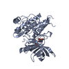
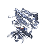
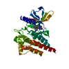
[English] 日本語
Yorodumi
- PDB-4hw7: Crystal structure of FMS kinase domain with a small molecular inh... -

+
Open data


ID or keywords:

Loading...

-
Basic information

Entry
Database: PDB / ID: 4hw7
TitleCrystal structure of FMS kinase domain with a small molecular inhibitor, PLX647-OME
ComponentsMacrophage colony-stimulating factor 1 receptor
KeywordsTRANSFERASE/TRANSFERASE INHIBITOR / CSF-1-R / FMS PROTO-ONCOGENE / C-FMS / CD115 antigen / Kinase / ATP-binding / TRANSFERASE-TRANSFERASE INHIBITOR complex
Function / homology
Function and homology information


macrophage colony-stimulating factor receptor activity / forebrain neuron differentiation / CSF1-CSF1R complex / macrophage colony-stimulating factor signaling pathway / cell-cell junction maintenance / regulation of macrophage migration / cellular response to macrophage colony-stimulating factor stimulus / microglial cell proliferation / olfactory bulb development / mammary gland duct morphogenesis ...macrophage colony-stimulating factor receptor activity / forebrain neuron differentiation / CSF1-CSF1R complex / macrophage colony-stimulating factor signaling pathway / cell-cell junction maintenance / regulation of macrophage migration / cellular response to macrophage colony-stimulating factor stimulus / microglial cell proliferation / olfactory bulb development / mammary gland duct morphogenesis / positive regulation by host of viral process / ruffle organization / positive regulation of macrophage proliferation / regulation of bone resorption / positive regulation of cell motility / Other interleukin signaling / positive regulation of macrophage chemotaxis / growth factor binding / cellular response to cytokine stimulus / cytokine binding / monocyte differentiation / regulation of MAPK cascade / macrophage differentiation / hemopoiesis / positive regulation of protein tyrosine kinase activity / Transcriptional Regulation by VENTX / positive regulation of chemokine production / positive regulation of tyrosine phosphorylation of STAT protein / cell surface receptor protein tyrosine kinase signaling pathway / osteoclast differentiation / axon guidance / response to ischemia / regulation of actin cytoskeleton organization / receptor protein-tyrosine kinase / cytokine-mediated signaling pathway / peptidyl-tyrosine phosphorylation / Signaling by CSF1 (M-CSF) in myeloid cells / regulation of cell shape / protein phosphatase binding / protein tyrosine kinase activity / cell population proliferation / protein autophosphorylation / positive regulation of ERK1 and ERK2 cascade / positive regulation of phosphatidylinositol 3-kinase/protein kinase B signal transduction / receptor complex / positive regulation of cell migration / inflammatory response / positive regulation of protein phosphorylation / negative regulation of cell population proliferation / intracellular membrane-bounded organelle / innate immune response / positive regulation of cell population proliferation / negative regulation of apoptotic process / cell surface / signal transduction / protein homodimerization activity / nucleoplasm / ATP binding / plasma membrane
Similarity search - Function
Macrophage colony-stimulating factor 1 receptor / Tyrosine-protein kinase, receptor class III, conserved site / Receptor tyrosine kinase class III signature. / Immunoglobulin / Immunoglobulin domain / Immunoglobulin domain / : / Immunoglobulin subtype 2 / Immunoglobulin C-2 Type / Tyrosine-protein kinase, catalytic domain ...Macrophage colony-stimulating factor 1 receptor / Tyrosine-protein kinase, receptor class III, conserved site / Receptor tyrosine kinase class III signature. / Immunoglobulin / Immunoglobulin domain / Immunoglobulin domain / : / Immunoglobulin subtype 2 / Immunoglobulin C-2 Type / Tyrosine-protein kinase, catalytic domain / Tyrosine kinase, catalytic domain / Tyrosine protein kinases specific active-site signature. / Immunoglobulin subtype / Immunoglobulin / Tyrosine-protein kinase, active site / Protein tyrosine and serine/threonine kinase / Serine-threonine/tyrosine-protein kinase, catalytic domain / Transferase(Phosphotransferase) domain 1 / Transferase(Phosphotransferase); domain 1 / Phosphorylase Kinase; domain 1 / Phosphorylase Kinase; domain 1 / Ig-like domain profile. / Immunoglobulin-like domain / Immunoglobulin-like domain superfamily / Protein kinase, ATP binding site / Protein kinases ATP-binding region signature. / Immunoglobulin-like fold / Protein kinase domain profile. / Protein kinase domain / Protein kinase-like domain superfamily / 2-Layer Sandwich / Orthogonal Bundle / Mainly Alpha / Alpha Beta
Similarity search - Domain/homology
Chem-64M / Macrophage colony-stimulating factor 1 receptor
Similarity search - Component
Biological speciesHomo sapiens (human)
MethodX-RAY DIFFRACTION / SYNCHROTRON / MOLECULAR REPLACEMENT / Resolution: 2.9001 Å
AuthorsZhang, Y. / Zhang, C.
CitationJournal: Proc.Natl.Acad.Sci.USA / Year: 2013
Title: Design and pharmacology of a highly specific dual FMS and KIT kinase inhibitor.
Authors: Zhang, C. / Ibrahim, P.N. / Zhang, J. / Burton, E.A. / Habets, G. / Zhang, Y. / Powell, B. / West, B.L. / Matusow, B. / Tsang, G. / Shellooe, R. / Carias, H. / Nguyen, H. / Marimuthu, A. / ...Authors: Zhang, C. / Ibrahim, P.N. / Zhang, J. / Burton, E.A. / Habets, G. / Zhang, Y. / Powell, B. / West, B.L. / Matusow, B. / Tsang, G. / Shellooe, R. / Carias, H. / Nguyen, H. / Marimuthu, A. / Zhang, K.Y. / Oh, A. / Bremer, R. / Hurt, C.R. / Artis, D.R. / Wu, G. / Nespi, M. / Spevak, W. / Lin, P. / Nolop, K. / Hirth, P. / Tesch, G.H. / Bollag, G.
History
DepositionNov 7, 2012Deposition site: RCSB / Processing site: RCSB
Revision 1.0Mar 27, 2013Provider: repository / Type: Initial release
Revision 1.1Apr 17, 2013Group: Database references
Revision 1.2Feb 28, 2024Group: Data collection / Database references / Derived calculations
Category: chem_comp_atom / chem_comp_bond ...chem_comp_atom / chem_comp_bond / database_2 / struct_ref_seq_dif / struct_site
Item: _database_2.pdbx_DOI / _database_2.pdbx_database_accession ..._database_2.pdbx_DOI / _database_2.pdbx_database_accession / _struct_ref_seq_dif.details / _struct_site.pdbx_auth_asym_id / _struct_site.pdbx_auth_comp_id / _struct_site.pdbx_auth_seq_id

-
Structure visualization

Structure viewerMolecule:
MolmilJmol/JSmol

Downloads & links

-
Assembly

Deposited unit
A: Macrophage colony-stimulating factor 1 receptor
hetero molecules


Theoretical massNumber of molelcules
Total (without water)39,0532
Polymers38,6401
Non-polymers4121
Water00
1


  • Idetical with deposited unit
  • defined by author
TypeNameSymmetry operationNumber
identity operation1_555x,y,z1
2
A: Macrophage colony-stimulating factor 1 receptor
hetero molecules

A: Macrophage colony-stimulating factor 1 receptor
hetero molecules


Theoretical massNumber of molelcules
Total (without water)78,1054
Polymers77,2802
Non-polymers8252
Water0
TypeNameSymmetry operationNumber
identity operation1_555x,y,z1
crystal symmetry operation7_556y,x,-z+11
Buried area1960 Å2
ΔGint-12 kcal/mol
Surface area26090 Å2
MethodPISA
Unit cell
Length a, b, c (Å)62.964, 62.964, 182.780
Angle α, β, γ (deg.)90.00, 90.00, 90.00
Int Tables number96
Space group name H-MP43212

-
Components

#1: Protein Macrophage colony-stimulating factor 1 receptor / CSF-1 receptor / CSF-1-R / CSF-1R / M-CSF-R / Proto-oncogene c-Fms


Mass: 38640.176 Da / Num. of mol.: 1 / Fragment: FMS kinase domain with KID / Mutation: C667T, C830S, C907T
Source method: isolated from a genetically manipulated source
Source: (gene. exp.) Homo sapiens (human) / Gene: CSF1R, FMS / Cell line (production host): SF9 / Production host: SPODOPTERA FRUGIPERDA (fall armyworm)
References: UniProt: P07333, receptor protein-tyrosine kinase
#2: Chemical ChemComp-64M / 5-[(5-methoxy-1H-pyrrolo[2,3-b]pyridin-3-yl)methyl]-N-[4-(trifluoromethyl)benzyl]pyridin-2-amine


Mass: 412.408 Da / Num. of mol.: 1 / Source method: obtained synthetically / Formula: C22H19F3N4O

-
Experimental details

-
Experiment

ExperimentMethod: X-RAY DIFFRACTION / Number of used crystals: 1

-
Sample preparation

CrystalDensity Matthews: 2.35 Å3/Da / Density % sol: 47.59 %
Crystal growTemperature: 277 K / Method: vapor diffusion, sitting drop / pH: 7.5
Details: 20% PEG8K, 0.2M MgCl and 0.1M Tris, pH 7.5, VAPOR DIFFUSION, SITTING DROP, temperature 277K

-
Data collection

DiffractionMean temperature: 100 K
Diffraction sourceSource: SYNCHROTRON / Site: ALS / Beamline: 8.3.1 / Wavelength: 1.1159 Å
DetectorType: ADSC QUANTUM 315r / Detector: CCD / Date: Aug 11, 2012
RadiationMonochromator: Double flat crystal, Si(111) / Protocol: SINGLE WAVELENGTH / Monochromatic (M) / Laue (L): M / Scattering type: x-ray
Radiation wavelengthWavelength: 1.1159 Å / Relative weight: 1
ReflectionResolution: 2.9→59.5 Å / Num. all: 8769 / Num. obs: 8735 / % possible obs: 99.6 % / Observed criterion σ(F): -3 / Observed criterion σ(I): -3
Reflection shellResolution: 2.9→3.06 Å / % possible all: 99.8

-
Processing

Software
NameVersionClassification
Blu-Icedata collection
MOLREPphasing
PHENIX(phenix.refine: 1.7.3_928)refinement
MOSFLMdata reduction
SCALAdata scaling
RefinementMethod to determine structure: MOLECULAR REPLACEMENT / Resolution: 2.9001→51.9 Å / SU ML: 0.52 / σ(F): 1.36 / Phase error: 26.97 / Stereochemistry target values: ML
RfactorNum. reflection% reflectionSelection details
Rfree0.267 414 4.75 %RANDOM
Rwork0.22 ---
all0.222 8769 --
obs0.222 8714 99.38 %-
Solvent computationShrinkage radii: 0.9 Å / VDW probe radii: 1.11 Å / Solvent model: FLAT BULK SOLVENT MODEL / Bsol: 40.606 Å2 / ksol: 0.357 e/Å3
Displacement parameters
Baniso -1Baniso -2Baniso -3
1-10.2841 Å20 Å2-0 Å2
2--10.2841 Å2-0 Å2
3----20.5683 Å2
Refinement stepCycle: LAST / Resolution: 2.9001→51.9 Å
ProteinNucleic acidLigandSolventTotal
Num. atoms2262 0 30 0 2292
Refine LS restraints
Refine-IDTypeDev idealNumber
X-RAY DIFFRACTIONf_bond_d0.0052349
X-RAY DIFFRACTIONf_angle_d0.8913183
X-RAY DIFFRACTIONf_dihedral_angle_d14.038847
X-RAY DIFFRACTIONf_chiral_restr0.057345
X-RAY DIFFRACTIONf_plane_restr0.005404
LS refinement shell
Resolution (Å)Rfactor RfreeNum. reflection RfreeRfactor RworkNum. reflection RworkRefine-ID% reflection obs (%)
2.9001-3.31960.38851370.29942690X-RAY DIFFRACTION99
3.3196-4.18210.27931350.21912728X-RAY DIFFRACTION100
4.1821-51.85790.21931420.1932882X-RAY DIFFRACTION99
Refinement TLS params.

Method: refined / Refine-ID: X-RAY DIFFRACTION

IDL112)L122)L132)L222)L232)L332)S11 (Å °)S12 (Å °)S13 (Å °)S21 (Å °)S22 (Å °)S23 (Å °)S31 (Å °)S32 (Å °)S33 (Å °)T112)T122)T132)T222)T232)T332)Origin x (Å)Origin y (Å)Origin z (Å)
11.5689-1.1011-1.06361.69570.21137.5040.134-0.06250.1119-0.0227-0.0516-0.17160.09890.3495-0.08890.2567-0.0402-0.06770.2945-0.01210.390636.412212.832282.4986
20.4707-0.16970.56060.2164-0.24640.68170.05850.03670.0222-0.08990.1604-0.0276-0.0112-0.1175-0.18371.05990.06730.11871.25250.23310.767336.503212.084874.8879
Refinement TLS group
IDRefine-IDRefine TLS-IDSelection details
1X-RAY DIFFRACTION1chain A and resid 566:914
2X-RAY DIFFRACTION2chain A and resid 1001:1001

+
About Yorodumi

-
News

-
Feb 9, 2022. New format data for meta-information of EMDB entries

New format data for meta-information of EMDB entries

  • Version 3 of the EMDB header file is now the official format.
  • The previous official version 1.9 will be removed from the archive.

Related info.:EMDB header

External links:wwPDB to switch to version 3 of the EMDB data model

-
Aug 12, 2020. Covid-19 info

Covid-19 info

URL: https://pdbjlc1.pdbj.org/emnavi/covid19.php

New page: Covid-19 featured information page in EM Navigator.

Related info.:Covid-19 info / Mar 5, 2020. Novel coronavirus structure data

+
Mar 5, 2020. Novel coronavirus structure data

Novel coronavirus structure data

Related info.:Yorodumi Speices / Aug 12, 2020. Covid-19 info

External links:COVID-19 featured content - PDBj / Molecule of the Month (242):Coronavirus Proteases

+
Jan 31, 2019. EMDB accession codes are about to change! (news from PDBe EMDB page)

EMDB accession codes are about to change! (news from PDBe EMDB page)

  • The allocation of 4 digits for EMDB accession codes will soon come to an end. Whilst these codes will remain in use, new EMDB accession codes will include an additional digit and will expand incrementally as the available range of codes is exhausted. The current 4-digit format prefixed with “EMD-” (i.e. EMD-XXXX) will advance to a 5-digit format (i.e. EMD-XXXXX), and so on. It is currently estimated that the 4-digit codes will be depleted around Spring 2019, at which point the 5-digit format will come into force.
  • The EM Navigator/Yorodumi systems omit the EMD- prefix.

Related info.:Q: What is EMD? / ID/Accession-code notation in Yorodumi/EM Navigator

External links:EMDB Accession Codes are Changing Soon! / Contact to PDBj

+
Jul 12, 2017. Major update of PDB

Major update of PDB

  • wwPDB released updated PDB data conforming to the new PDBx/mmCIF dictionary.
  • This is a major update changing the version number from 4 to 5, and with Remediation, in which all the entries are updated.
  • In this update, many items about electron microscopy experimental information are reorganized (e.g. em_software).
  • Now, EM Navigator and Yorodumi are based on the updated data.

External links:wwPDB Remediation / Enriched Model Files Conforming to OneDep Data Standards Now Available in the PDB FTP Archive

-
Yorodumi

Thousand views of thousand structures

  • Yorodumi is a browser for structure data from EMDB, PDB, SASBDB, etc.
  • This page is also the successor to EM Navigator detail page, and also detail information page/front-end page for Omokage search.
  • The word "yorodu" (or yorozu) is an old Japanese word meaning "ten thousand". "mi" (miru) is to see.

Related info.:EMDB / PDB / SASBDB / Comparison of 3 databanks / Yorodumi Search / Aug 31, 2016. New EM Navigator & Yorodumi / Yorodumi Papers / Jmol/JSmol / Function and homology information / Changes in new EM Navigator and Yorodumi

Read more