 Movie
Movie Controller

[English] 日本語
 Yorodumi
- PDB-4hll: Crystal structure of Artificial ankyrin repeat protein_Ank(GAG)1D4 -
+ Open data

- Basic information
| Entry | Database: PDB / ID: 4hll | ||||||
|---|---|---|---|---|---|---|---|
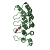
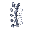
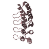
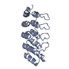
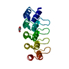
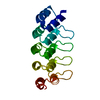
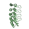
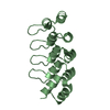
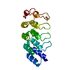
| Title | Crystal structure of Artificial ankyrin repeat protein_Ank(GAG)1D4 | ||||||
 Components Components | Ankyrin(GAG)1D4 | ||||||
 Keywords Keywords | PROTEIN BINDING / Ankyrin repeats | ||||||
| Function / homology | Ankyrin repeat-containing domain / Serine Threonine Protein Phosphatase 5, Tetratricopeptide repeat / Alpha Horseshoe / Mainly Alpha Function and homology information Function and homology information | ||||||
| Biological species |  | ||||||
| Method |  X-RAY DIFFRACTION / SYNCHROTRON / MOLECULAR REPLACEMENT / Resolution: 2.2 Å X-RAY DIFFRACTION / SYNCHROTRON / MOLECULAR REPLACEMENT / Resolution: 2.2 Å | ||||||
 Authors Authors | Chuankhayan, P. / Nangola, S. / Minard, P. / Boulanger, P. / Hong, S.S. / Tayapiwatana, C. / Chen, C.-J. | ||||||
 Citation Citation | Journal: To be Published Title: Identification of Gag bioactive determinants specific to designed ankyrin and interfering in HIV-1 assembly Authors: Chuankhayan, P. / Nangola, S. / Minard, P. / Boulanger, P. / Hong, S.S. / Tayapiwatana, C. / Chen, C.-J. | ||||||
| History |
|
- Structure visualization
| Structure viewer | Molecule: MolmilJmol/JSmol |
|---|
- Downloads & links
-Download
| PDBx/mmCIF format | 4hll.cif.gz | 41.9 KB | Display | PDBx/mmCIF format |
|---|---|---|---|---|
| PDB format | pdb4hll.ent.gz | 28.7 KB | Display | PDB format |
| PDBx/mmJSON format | 4hll.json.gz | Tree view | PDBx/mmJSON format | |
| Others |  Other downloads Other downloads |
-Validation report
| Arichive directory | https://data.pdbj.org/pub/pdb/validation_reports/hl/4hllftp://data.pdbj.org/pub/pdb/validation_reports/hl/4hll | HTTPS FTP |
|---|
-Related structure data
| Related structure data | 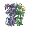 3nocS S: Starting model for refinement |
|---|---|
| Similar structure data |


-Links
 PDBj
PDBj- Assembly
| Deposited unit | 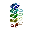
| ||||||||
|---|---|---|---|---|---|---|---|---|---|
| 1 |
| ||||||||
| Unit cell |
|
-Components
| #1: Protein | Mass: 18777.041 Da / Num. of mol.: 1 Source method: isolated from a genetically manipulated source Source: (gene. exp.) Description: THE SEQUENCE DERIVED FROM A PHAGE-DISPLAYED LIBRARY PRODUCED FOR E.COLI. Plasmid: pQE30 / Production host: |
|---|---|
| #2: Water | ChemComp-HOH /  Mass: 18.015 Da / Num. of mol.: 34 / Source method: isolated from a natural source / Formula: H2O Mass: 18.015 Da / Num. of mol.: 34 / Source method: isolated from a natural source / Formula: H2O |
| Sequence details | THE SEQUENCE OF THIS PROTEIN WAS NOT AVAILABLE AT THE UNIPROT KNOWLEDGEBASE DATABASE (UNIPROTKB) AT ...THE SEQUENCE OF THIS PROTEIN WAS NOT AVAILABLE AT THE UNIPROT KNOWLEDGEB |
-Experimental details
-Experiment
| Experiment | Method: X-RAY DIFFRACTION / Number of used crystals: 1 |
|---|
- Sample preparation
| Crystal | Density Matthews: 2.07 Å3/Da / Density % sol: 40.64 % |
|---|---|
| Crystal grow | Temperature: 291 K / Method: vapor diffusion, hanging drop / pH: 7.5 Details: 25% PEG 400, 5% PEG 3000, 10% glycerol, 0.1M Hepes, pH 7.5, VAPOR DIFFUSION, HANGING DROP, temperature 291K |
-Data collection
| Diffraction |
| |||||||||||||||
|---|---|---|---|---|---|---|---|---|---|---|---|---|---|---|---|---|
| Diffraction source |
| |||||||||||||||
| Detector |
| |||||||||||||||
| Radiation | Monochromator: Si 111 CHANNEL / Protocol: SINGLE WAVELENGTH / Monochromatic (M) / Laue (L): M / Scattering type: x-ray | |||||||||||||||
| Radiation wavelength | Wavelength: 1 Å / Relative weight: 1 | |||||||||||||||
| Reflection | Resolution: 2.2→30 Å / Num. obs: 8038 / % possible obs: 99.3 % / Observed criterion σ(F): 0 / Observed criterion σ(I): 0 / Redundancy: 3.4 % / Rsym value: 0.049 | |||||||||||||||
| Reflection shell | Resolution: 2.2→2.3 Å / % possible all: 99.6 |


- Processing
| Software |
| ||||||||||||||||||||
|---|---|---|---|---|---|---|---|---|---|---|---|---|---|---|---|---|---|---|---|---|---|
| Refinement | Method to determine structure: MOLECULAR REPLACEMENT Starting model: 3NOC Resolution: 2.2→30 Å / Occupancy max: 1 / Occupancy min: 1 / Cross valid method: THROUGHOUT / σ(F): 0 / Stereochemistry target values: Engh & Huber
| ||||||||||||||||||||
| Solvent computation | Bsol: 67.432 Å2 | ||||||||||||||||||||
| Displacement parameters | Biso max: 134.65 Å2 / Biso mean: 36.5881 Å2 / Biso min: 14.5 Å2
| ||||||||||||||||||||
| Refinement step | Cycle: LAST / Resolution: 2.2→30 Å
| ||||||||||||||||||||
| Refine LS restraints |
| ||||||||||||||||||||
| LS refinement shell | Resolution: 2.2→2.3 Å | ||||||||||||||||||||
| Xplor file |
|
