 Movie
Movie Controller

[English] 日本語
 Yorodumi
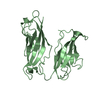
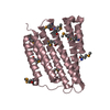
- PDB-4hi1: Crystal structure of acylphosphatase C20R mutant from Vibrio chol... -
+ Open data

- Basic information
| Entry | Database: PDB / ID: 4hi1 | ||||||
|---|---|---|---|---|---|---|---|
| Title | Crystal structure of acylphosphatase C20R mutant from Vibrio cholerae0395 | ||||||
 Components Components | Acylphosphatase | ||||||
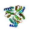
 Keywords Keywords | HYDROLASE / Ferredoxin fold | ||||||
| Function / homology |  Function and homology information Function and homology information | ||||||
| Biological species |  Vibrio cholerae O395 (bacteria) Vibrio cholerae O395 (bacteria) | ||||||
| Method |  X-RAY DIFFRACTION / MOLECULAR REPLACEMENT / Resolution: 1.964 Å X-RAY DIFFRACTION / MOLECULAR REPLACEMENT / Resolution: 1.964 Å | ||||||
 Authors Authors | Nath, S. / Banerjee, R. / Sen, U. | ||||||
 Citation Citation | Journal: J.Mol.Biol. / Year: 2013 Title: Crystal structure of acylphosphatase C20R mutant from Vibrio cholerae0395 Authors: Nath, S. / Banerjee, R. / Sen, U. | ||||||
| History |
|
- Structure visualization
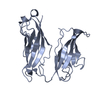
| Structure viewer | Molecule: MolmilJmol/JSmol |
|---|
- Downloads & links
-Download
| PDBx/mmCIF format | 4hi1.cif.gz | 124.7 KB | Display | PDBx/mmCIF format |
|---|---|---|---|---|
| PDB format | pdb4hi1.ent.gz | 98.7 KB | Display | PDB format |
| PDBx/mmJSON format | 4hi1.json.gz | Tree view | PDBx/mmJSON format | |
| Others |  Other downloads Other downloads |
-Validation report
| Arichive directory | https://data.pdbj.org/pub/pdb/validation_reports/hi/4hi1ftp://data.pdbj.org/pub/pdb/validation_reports/hi/4hi1 | HTTPS FTP |
|---|
-Related structure data
| Related structure data |  4hi2C  2bjdS C: citing same article ( S: Starting model for refinement |
|---|---|
| Similar structure data |


-Links
 PDBj
PDBj- Assembly
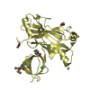
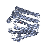
| Deposited unit | 
| ||||||||
|---|---|---|---|---|---|---|---|---|---|
| 1 |
| ||||||||
| Unit cell |
|
-Components
| #1: Protein | Mass: 10292.619 Da / Num. of mol.: 3 / Mutation: C20R Source method: isolated from a genetically manipulated source Source: (gene. exp.) Vibrio cholerae O395 (bacteria) / Strain: ATCC 39541 / Ogawa 395 / O395 / Gene: acyP, VC0395_A0969, VC395_1474 / Plasmid: pET28a+ / Production host: #2: Chemical |   Mass: 159.938 Da / Num. of mol.: 3 / Source method: obtained synthetically / Formula: MoO4 Mass: 159.938 Da / Num. of mol.: 3 / Source method: obtained synthetically / Formula: MoO4#3: Chemical |   Mass: 96.063 Da / Num. of mol.: 3 / Source method: obtained synthetically / Formula: SO4 Mass: 96.063 Da / Num. of mol.: 3 / Source method: obtained synthetically / Formula: SO4#4: Water | ChemComp-HOH / |  Mass: 18.015 Da / Num. of mol.: 249 / Source method: isolated from a natural source / Formula: H2O Mass: 18.015 Da / Num. of mol.: 249 / Source method: isolated from a natural source / Formula: H2OHas protein modification | Y | |
|---|
-Experimental details
-Experiment
| Experiment | Method: X-RAY DIFFRACTION / Number of used crystals: 1 |
|---|
- Sample preparation
| Crystal | Density Matthews: 2.12 Å3/Da / Density % sol: 41.95 % |
|---|---|
| Crystal grow | Temperature: 293 K / Method: vapor diffusion, hanging drop / pH: 9 Details: 1.6M ammonium sulfate, 0.1M bicine pH 9.0, VAPOR DIFFUSION, HANGING DROP, temperature 293K |
-Data collection
| Diffraction | Mean temperature: 100 K |
|---|---|
| Diffraction source | Source: ROTATING ANODE / Type: ENRAF-NONIUS FR591 / Wavelength: 1.5418 Å |
| Detector | Type: MAR scanner 345 mm plate / Detector: IMAGE PLATE / Date: Jul 13, 2012 |
| Radiation | Monochromator: Ni FILTER / Protocol: SINGLE WAVELENGTH / Monochromatic (M) / Laue (L): M / Scattering type: x-ray |
| Radiation wavelength | Wavelength: 1.5418 Å / Relative weight: 1 |
| Reflection | Resolution: 1.964→39.708 Å / Num. obs: 17237 / % possible obs: 93.5 % |
| Reflection shell | Resolution: 1.96→2.06 Å / Redundancy: 3.8 % / Rmerge(I) obs: 0.062 / Mean I/σ(I) obs: 12.5 / Num. unique all: 17237 / % possible all: 93.5 |
- Processing
| Software |
| |||||||||||||||||||||||||||||||||||||||||||||||||||||||||||||||||||||||||||||||||||||||||||
|---|---|---|---|---|---|---|---|---|---|---|---|---|---|---|---|---|---|---|---|---|---|---|---|---|---|---|---|---|---|---|---|---|---|---|---|---|---|---|---|---|---|---|---|---|---|---|---|---|---|---|---|---|---|---|---|---|---|---|---|---|---|---|---|---|---|---|---|---|---|---|---|---|---|---|---|---|---|---|---|---|---|---|---|---|---|---|---|---|---|---|---|---|
| Refinement | Method to determine structure: MOLECULAR REPLACEMENT Starting model: 2BJD Resolution: 1.964→39.708 Å / SU ML: 0.23 / Phase error: 26.54 / Stereochemistry target values: ML
| |||||||||||||||||||||||||||||||||||||||||||||||||||||||||||||||||||||||||||||||||||||||||||
| Solvent computation | Shrinkage radii: 0.9 Å / VDW probe radii: 1.11 Å / Solvent model: FLAT BULK SOLVENT MODEL | |||||||||||||||||||||||||||||||||||||||||||||||||||||||||||||||||||||||||||||||||||||||||||
| Refine analyze | Luzzati sigma a obs: 0.23 Å | |||||||||||||||||||||||||||||||||||||||||||||||||||||||||||||||||||||||||||||||||||||||||||
| Refinement step | Cycle: LAST / Resolution: 1.964→39.708 Å
| |||||||||||||||||||||||||||||||||||||||||||||||||||||||||||||||||||||||||||||||||||||||||||
| Refine LS restraints |
| |||||||||||||||||||||||||||||||||||||||||||||||||||||||||||||||||||||||||||||||||||||||||||
| LS refinement shell |
| |||||||||||||||||||||||||||||||||||||||||||||||||||||||||||||||||||||||||||||||||||||||||||
| Refinement TLS params. | Method: refined / Origin x: 34.199 Å / Origin y: 27.7704 Å / Origin z: 20.3423 Å
| |||||||||||||||||||||||||||||||||||||||||||||||||||||||||||||||||||||||||||||||||||||||||||
| Refinement TLS group | Selection details: all |
