chloroplast avoidance movement / chloroplast accumulation movement / cellular response to blue light / leaf phyllotactic patterning / regulation of proton transport / response to red or far red light / regulation of leaf morphogenesis / phototropism / blue light signaling pathway / regulation of stomatal movement ...chloroplast avoidance movement / chloroplast accumulation movement / cellular response to blue light / leaf phyllotactic patterning / regulation of proton transport / response to red or far red light / regulation of leaf morphogenesis / phototropism / blue light signaling pathway / regulation of stomatal movement / blue light photoreceptor activity / response to blue light / plant-type vacuole / circadian rhythm / cytoplasmic side of plasma membrane / kinase activity / FMN binding / protein kinase activity / non-specific serine/threonine protein kinase / protein serine kinase activity / protein serine/threonine kinase activity / mRNA binding / cell surface / ATP binding / identical protein binding / nucleus / plasma membrane / cytoplasm Similarity search - Function
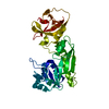
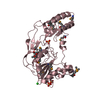
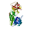
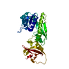
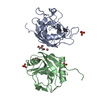
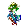
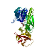
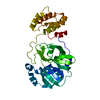
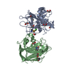
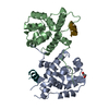
PAS domain / PAS-associated, C-terminal / PAC domain profile. / PAC motif / Motif C-terminal to PAS motifs (likely to contribute to PAS structural domain) / PAS domain / Beta-Lactamase / PAS domain / PAS repeat profile. / PAS domain ...PAS domain / PAS-associated, C-terminal / PAC domain profile. / PAC motif / Motif C-terminal to PAS motifs (likely to contribute to PAS structural domain) / PAS domain / Beta-Lactamase / PAS domain / PAS repeat profile. / PAS domain / PAS domain superfamily / Serine/threonine-protein kinase, active site / Serine/Threonine protein kinases active-site signature. / Protein kinase domain / Serine/Threonine protein kinases, catalytic domain / Protein kinase, ATP binding site / Protein kinases ATP-binding region signature. / Protein kinase domain profile. / Protein kinase domain / Protein kinase-like domain superfamily / 2-Layer Sandwich / Alpha Beta Similarity search - Domain/homology
Mass: 18.015 Da / Num. of mol.: 29 / Source method: isolated from a natural source / Formula: H2O
-
Experimental details
-
Experiment
Experiment
Method: X-RAY DIFFRACTION / Number of used crystals: 1
-
Sample preparation
Crystal
Density Matthews: 2.25 Å3/Da / Density % sol: 45.33 %
Crystal grow
Temperature: 287 K / Method: vapor diffusion, sitting drop / pH: 7.4 Details: Protein at 21 mg/mL in 20 mM TRIS-HCl pH 7.4, 20 mM NaCl, 15 % glycerol Crystallization: The JCSG+ Suite (D12: 0.04 M Potassium phosphate, 16% (w/v) PEG 8000, 20% (v/v) glycerol), VAPOR ...Details: Protein at 21 mg/mL in 20 mM TRIS-HCl pH 7.4, 20 mM NaCl, 15 % glycerol Crystallization: The JCSG+ Suite (D12: 0.04 M Potassium phosphate, 16% (w/v) PEG 8000, 20% (v/v) glycerol), VAPOR DIFFUSION, SITTING DROP, temperature 287K
In the structure databanks used in Yorodumi, some data are registered as the other names, "COVID-19 virus" and "2019-nCoV". Here are the details of the virus and the list of structure data.
Jan 31, 2019. EMDB accession codes are about to change! (news from PDBe EMDB page)
EMDB accession codes are about to change! (news from PDBe EMDB page)
The allocation of 4 digits for EMDB accession codes will soon come to an end. Whilst these codes will remain in use, new EMDB accession codes will include an additional digit and will expand incrementally as the available range of codes is exhausted. The current 4-digit format prefixed with “EMD-” (i.e. EMD-XXXX) will advance to a 5-digit format (i.e. EMD-XXXXX), and so on. It is currently estimated that the 4-digit codes will be depleted around Spring 2019, at which point the 5-digit format will come into force.
The EM Navigator/Yorodumi systems omit the EMD- prefix.
Related info.:Q: What is EMD? / ID/Accession-code notation in Yorodumi/EM Navigator
Yorodumi is a browser for structure data from EMDB, PDB, SASBDB, etc.
This page is also the successor to EM Navigator detail page, and also detail information page/front-end page for Omokage search.
The word "yorodu" (or yorozu) is an old Japanese word meaning "ten thousand". "mi" (miru) is to see.
Related info.:EMDB / PDB / SASBDB / Comparison of 3 databanks / Yorodumi Search / Aug 31, 2016. New EM Navigator & Yorodumi / Yorodumi Papers / Jmol/JSmol / Function and homology information / Changes in new EM Navigator and Yorodumi
 Movie
Movie Controller

 Yorodumi
 Open data
 Basic information
 Components
 Keywords
 Function and homology information
 X-RAY DIFFRACTION /
  Authors
 Citation
 Structure visualization
 Downloads & links
 Other downloads
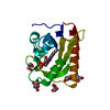
 PDBj
PDBj


 Assembly
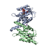


 Mass: 456.344 Da / Num. of mol.: 2 / Source method: obtained synthetically / Formula: C17H21N4O9P
 Mass: 22.990 Da / Num. of mol.: 1 / Source method: obtained synthetically / Formula: Na
 Mass: 18.015 Da / Num. of mol.: 29 / Source method: isolated from a natural source / Formula: H2O
 Sample preparation
 / Beamline: 14-BM-C / Wavelength: 0.9002 Å
 Processing